Tarapong Srisongkram*, Nur Fadhilah Syahid, Thanawat Piyasawetkul, Pannaphat Thirawatthanasak, Patcharapa Khamtang, Nathida Sawasnopparat, Dheerapat Tookkane, Natthida Weerapreeyakul and Ploenthip Puthongking,
{"title":"利用荧光染色和卷积神经网络预测球形细胞死亡","authors":"Tarapong Srisongkram*, Nur Fadhilah Syahid, Thanawat Piyasawetkul, Pannaphat Thirawatthanasak, Patcharapa Khamtang, Nathida Sawasnopparat, Dheerapat Tookkane, Natthida Weerapreeyakul and Ploenthip Puthongking, ","doi":"10.1021/acs.chemrestox.3c00257","DOIUrl":null,"url":null,"abstract":"<p >Three-dimensional (3D) cell culture is emerging for drug design and drug screening. Skin toxicity is one of the most important assays for determining the toxicity of a compound before being used in skin application. Much work has been done to find an alternative assay without animal experiments. 3D cell culture is one of the methods that provides clinically relevant models with superior clinical translation compared to that of 2D cell culture. In this study, we developed a spheroid toxicity assay using keratinocyte HaCaT cells with propidium iodide and calcein AM. We also applied the transfer learning-containing convolutional neural network (CNN) to further determine spheroid cell death with fluorescence labeling. Our result shows that the morphologies of the spheroid are the key features in determining the apoptosis cell death of the HaCaT spheroid. Our CNN model provided good statistical measurement in terms of accuracy, precision, and recall in both validation and external test data sets. One can predict keratinocyte spheroid cell death if that spheroid image contains the fluorescence signals from propidium iodide and calcein AM. The CNN model can be accessed in the web application at https://qsarlabs.com/#spheroiddeath.</p>","PeriodicalId":31,"journal":{"name":"Chemical Research in Toxicology","volume":"36 12","pages":"1980–1989"},"PeriodicalIF":3.8000,"publicationDate":"2023-12-05","publicationTypes":"Journal Article","fieldsOfStudy":null,"isOpenAccess":false,"openAccessPdf":"","citationCount":"0","resultStr":"{\"title\":\"Prediction of Spheroid Cell Death Using Fluorescence Staining and Convolutional Neural Networks\",\"authors\":\"Tarapong Srisongkram*, Nur Fadhilah Syahid, Thanawat Piyasawetkul, Pannaphat Thirawatthanasak, Patcharapa Khamtang, Nathida Sawasnopparat, Dheerapat Tookkane, Natthida Weerapreeyakul and Ploenthip Puthongking, \",\"doi\":\"10.1021/acs.chemrestox.3c00257\",\"DOIUrl\":null,\"url\":null,\"abstract\":\"<p >Three-dimensional (3D) cell culture is emerging for drug design and drug screening. Skin toxicity is one of the most important assays for determining the toxicity of a compound before being used in skin application. Much work has been done to find an alternative assay without animal experiments. 3D cell culture is one of the methods that provides clinically relevant models with superior clinical translation compared to that of 2D cell culture. In this study, we developed a spheroid toxicity assay using keratinocyte HaCaT cells with propidium iodide and calcein AM. We also applied the transfer learning-containing convolutional neural network (CNN) to further determine spheroid cell death with fluorescence labeling. Our result shows that the morphologies of the spheroid are the key features in determining the apoptosis cell death of the HaCaT spheroid. Our CNN model provided good statistical measurement in terms of accuracy, precision, and recall in both validation and external test data sets. One can predict keratinocyte spheroid cell death if that spheroid image contains the fluorescence signals from propidium iodide and calcein AM. The CNN model can be accessed in the web application at https://qsarlabs.com/#spheroiddeath.</p>\",\"PeriodicalId\":31,\"journal\":{\"name\":\"Chemical Research in Toxicology\",\"volume\":\"36 12\",\"pages\":\"1980–1989\"},\"PeriodicalIF\":3.8000,\"publicationDate\":\"2023-12-05\",\"publicationTypes\":\"Journal Article\",\"fieldsOfStudy\":null,\"isOpenAccess\":false,\"openAccessPdf\":\"\",\"citationCount\":\"0\",\"resultStr\":null,\"platform\":\"Semanticscholar\",\"paperid\":null,\"PeriodicalName\":\"Chemical Research in Toxicology\",\"FirstCategoryId\":\"3\",\"ListUrlMain\":\"https://pubs.acs.org/doi/10.1021/acs.chemrestox.3c00257\",\"RegionNum\":3,\"RegionCategory\":\"医学\",\"ArticlePicture\":[],\"TitleCN\":null,\"AbstractTextCN\":null,\"PMCID\":null,\"EPubDate\":\"\",\"PubModel\":\"\",\"JCR\":\"Q2\",\"JCRName\":\"CHEMISTRY, MEDICINAL\",\"Score\":null,\"Total\":0}","platform":"Semanticscholar","paperid":null,"PeriodicalName":"Chemical Research in Toxicology","FirstCategoryId":"3","ListUrlMain":"https://pubs.acs.org/doi/10.1021/acs.chemrestox.3c00257","RegionNum":3,"RegionCategory":"医学","ArticlePicture":[],"TitleCN":null,"AbstractTextCN":null,"PMCID":null,"EPubDate":"","PubModel":"","JCR":"Q2","JCRName":"CHEMISTRY, MEDICINAL","Score":null,"Total":0}
引用次数: 0
摘要
三维(3D)细胞培养正在成为药物设计和药物筛选的新兴手段。皮肤毒性是确定化合物在皮肤应用前毒性的最重要检测方法之一。为了找到一种无需动物实验的替代检测方法,人们做了大量工作。三维细胞培养是提供临床相关模型的方法之一,与二维细胞培养相比,三维细胞培养具有更好的临床转化效果。在本研究中,我们使用角质形成细胞 HaCaT 细胞与碘化丙啶和钙蓝蛋白 AM 共同开发了一种球形毒性检测方法。我们还应用了包含传递学习的卷积神经网络(CNN),通过荧光标记进一步确定球形细胞的死亡。结果表明,球形细胞的形态是确定 HaCaT 球形细胞凋亡的关键特征。我们的 CNN 模型在验证数据集和外部测试数据集的准确度、精确度和召回率方面都提供了良好的统计测量。如果球形图像包含碘化丙啶和钙蓝蛋白 AM 的荧光信号,就可以预测角质形成细胞球形体细胞死亡。CNN 模型可在 https://qsarlabs.com/#spheroiddeath 的网络应用程序中访问。
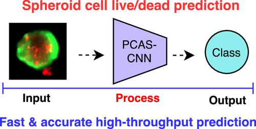
Prediction of Spheroid Cell Death Using Fluorescence Staining and Convolutional Neural Networks
Three-dimensional (3D) cell culture is emerging for drug design and drug screening. Skin toxicity is one of the most important assays for determining the toxicity of a compound before being used in skin application. Much work has been done to find an alternative assay without animal experiments. 3D cell culture is one of the methods that provides clinically relevant models with superior clinical translation compared to that of 2D cell culture. In this study, we developed a spheroid toxicity assay using keratinocyte HaCaT cells with propidium iodide and calcein AM. We also applied the transfer learning-containing convolutional neural network (CNN) to further determine spheroid cell death with fluorescence labeling. Our result shows that the morphologies of the spheroid are the key features in determining the apoptosis cell death of the HaCaT spheroid. Our CNN model provided good statistical measurement in terms of accuracy, precision, and recall in both validation and external test data sets. One can predict keratinocyte spheroid cell death if that spheroid image contains the fluorescence signals from propidium iodide and calcein AM. The CNN model can be accessed in the web application at https://qsarlabs.com/#spheroiddeath.
期刊介绍:
Chemical Research in Toxicology publishes Articles, Rapid Reports, Chemical Profiles, Reviews, Perspectives, Letters to the Editor, and ToxWatch on a wide range of topics in Toxicology that inform a chemical and molecular understanding and capacity to predict biological outcomes on the basis of structures and processes. The overarching goal of activities reported in the Journal are to provide knowledge and innovative approaches needed to promote intelligent solutions for human safety and ecosystem preservation. The journal emphasizes insight concerning mechanisms of toxicity over phenomenological observations. It upholds rigorous chemical, physical and mathematical standards for characterization and application of modern techniques.

 求助内容:
求助内容: 应助结果提醒方式:


