{"title":"基于模型的深度学习重建头颈部评估弥散加权成像图像质量改进","authors":"Noriyuki Fujima, Junichi Nakagawa, Hiroyuki Kameda, Yohei Ikebe, Taisuke Harada, Yukie Shimizu, Nayuta Tsushima, Satoshi Kano, Akihiro Homma, Jihun Kwon, Masami Yoneyama, Kohsuke Kudo","doi":"10.1007/s10334-023-01129-4","DOIUrl":null,"url":null,"abstract":"<p><strong>Objectives: </strong>To investigate the utility of deep learning (DL)-based image reconstruction using a model-based approach in head and neck diffusion-weighted imaging (DWI).</p><p><strong>Materials and methods: </strong>We retrospectively analyzed the cases of 41 patients who underwent head/neck DWI. The DWI in 25 patients demonstrated an untreated lesion. We performed qualitative and quantitative assessments in the DWI analyses with both deep learning (DL)- and conventional parallel imaging (PI)-based reconstructions. For the qualitative assessment, we visually evaluated the overall image quality, soft tissue conspicuity, degree of artifact(s), and lesion conspicuity based on a five-point system. In the quantitative assessment, we measured the signal-to-noise ratio (SNR) of the bilateral parotid glands, submandibular gland, the posterior muscle, and the lesion. We then calculated the contrast-to-noise ratio (CNR) between the lesion and the adjacent muscle.</p><p><strong>Results: </strong>Significant differences were observed in the qualitative analysis between the DWI with PI-based and DL-based reconstructions for all of the evaluation items (p < 0.001). In the quantitative analysis, significant differences in the SNR and CNR between the DWI with PI-based and DL-based reconstructions were observed for all of the evaluation items (p = 0.002 ~ p < 0.001).</p><p><strong>Discussion: </strong>DL-based image reconstruction with the model-based technique effectively provided sufficient image quality in head/neck DWI.</p>","PeriodicalId":18067,"journal":{"name":"Magnetic Resonance Materials in Physics, Biology and Medicine","volume":" ","pages":"439-447"},"PeriodicalIF":2.0000,"publicationDate":"2024-07-01","publicationTypes":"Journal Article","fieldsOfStudy":null,"isOpenAccess":false,"openAccessPdf":"","citationCount":"0","resultStr":"{\"title\":\"Improvement of image quality in diffusion-weighted imaging with model-based deep learning reconstruction for evaluations of the head and neck.\",\"authors\":\"Noriyuki Fujima, Junichi Nakagawa, Hiroyuki Kameda, Yohei Ikebe, Taisuke Harada, Yukie Shimizu, Nayuta Tsushima, Satoshi Kano, Akihiro Homma, Jihun Kwon, Masami Yoneyama, Kohsuke Kudo\",\"doi\":\"10.1007/s10334-023-01129-4\",\"DOIUrl\":null,\"url\":null,\"abstract\":\"<p><strong>Objectives: </strong>To investigate the utility of deep learning (DL)-based image reconstruction using a model-based approach in head and neck diffusion-weighted imaging (DWI).</p><p><strong>Materials and methods: </strong>We retrospectively analyzed the cases of 41 patients who underwent head/neck DWI. The DWI in 25 patients demonstrated an untreated lesion. We performed qualitative and quantitative assessments in the DWI analyses with both deep learning (DL)- and conventional parallel imaging (PI)-based reconstructions. For the qualitative assessment, we visually evaluated the overall image quality, soft tissue conspicuity, degree of artifact(s), and lesion conspicuity based on a five-point system. In the quantitative assessment, we measured the signal-to-noise ratio (SNR) of the bilateral parotid glands, submandibular gland, the posterior muscle, and the lesion. We then calculated the contrast-to-noise ratio (CNR) between the lesion and the adjacent muscle.</p><p><strong>Results: </strong>Significant differences were observed in the qualitative analysis between the DWI with PI-based and DL-based reconstructions for all of the evaluation items (p < 0.001). In the quantitative analysis, significant differences in the SNR and CNR between the DWI with PI-based and DL-based reconstructions were observed for all of the evaluation items (p = 0.002 ~ p < 0.001).</p><p><strong>Discussion: </strong>DL-based image reconstruction with the model-based technique effectively provided sufficient image quality in head/neck DWI.</p>\",\"PeriodicalId\":18067,\"journal\":{\"name\":\"Magnetic Resonance Materials in Physics, Biology and Medicine\",\"volume\":\" \",\"pages\":\"439-447\"},\"PeriodicalIF\":2.0000,\"publicationDate\":\"2024-07-01\",\"publicationTypes\":\"Journal Article\",\"fieldsOfStudy\":null,\"isOpenAccess\":false,\"openAccessPdf\":\"\",\"citationCount\":\"0\",\"resultStr\":null,\"platform\":\"Semanticscholar\",\"paperid\":null,\"PeriodicalName\":\"Magnetic Resonance Materials in Physics, Biology and Medicine\",\"FirstCategoryId\":\"3\",\"ListUrlMain\":\"https://doi.org/10.1007/s10334-023-01129-4\",\"RegionNum\":4,\"RegionCategory\":\"医学\",\"ArticlePicture\":[],\"TitleCN\":null,\"AbstractTextCN\":null,\"PMCID\":null,\"EPubDate\":\"2023/11/21 0:00:00\",\"PubModel\":\"Epub\",\"JCR\":\"Q3\",\"JCRName\":\"RADIOLOGY, NUCLEAR MEDICINE & MEDICAL IMAGING\",\"Score\":null,\"Total\":0}","platform":"Semanticscholar","paperid":null,"PeriodicalName":"Magnetic Resonance Materials in Physics, Biology and Medicine","FirstCategoryId":"3","ListUrlMain":"https://doi.org/10.1007/s10334-023-01129-4","RegionNum":4,"RegionCategory":"医学","ArticlePicture":[],"TitleCN":null,"AbstractTextCN":null,"PMCID":null,"EPubDate":"2023/11/21 0:00:00","PubModel":"Epub","JCR":"Q3","JCRName":"RADIOLOGY, NUCLEAR MEDICINE & MEDICAL IMAGING","Score":null,"Total":0}
Improvement of image quality in diffusion-weighted imaging with model-based deep learning reconstruction for evaluations of the head and neck.
Objectives: To investigate the utility of deep learning (DL)-based image reconstruction using a model-based approach in head and neck diffusion-weighted imaging (DWI).
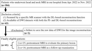
Materials and methods: We retrospectively analyzed the cases of 41 patients who underwent head/neck DWI. The DWI in 25 patients demonstrated an untreated lesion. We performed qualitative and quantitative assessments in the DWI analyses with both deep learning (DL)- and conventional parallel imaging (PI)-based reconstructions. For the qualitative assessment, we visually evaluated the overall image quality, soft tissue conspicuity, degree of artifact(s), and lesion conspicuity based on a five-point system. In the quantitative assessment, we measured the signal-to-noise ratio (SNR) of the bilateral parotid glands, submandibular gland, the posterior muscle, and the lesion. We then calculated the contrast-to-noise ratio (CNR) between the lesion and the adjacent muscle.
Results: Significant differences were observed in the qualitative analysis between the DWI with PI-based and DL-based reconstructions for all of the evaluation items (p < 0.001). In the quantitative analysis, significant differences in the SNR and CNR between the DWI with PI-based and DL-based reconstructions were observed for all of the evaluation items (p = 0.002 ~ p < 0.001).
Discussion: DL-based image reconstruction with the model-based technique effectively provided sufficient image quality in head/neck DWI.
期刊介绍:
MAGMA is a multidisciplinary international journal devoted to the publication of articles on all aspects of magnetic resonance techniques and their applications in medicine and biology. MAGMA currently publishes research papers, reviews, letters to the editor, and commentaries, six times a year. The subject areas covered by MAGMA include:
advances in materials, hardware and software in magnetic resonance technology,
new developments and results in research and practical applications of magnetic resonance imaging and spectroscopy related to biology and medicine,
study of animal models and intact cells using magnetic resonance,
reports of clinical trials on humans and clinical validation of magnetic resonance protocols.

 求助内容:
求助内容: 应助结果提醒方式:


