{"title":"基于压缩svd的L + S模型并行重构欠采样动态MRI数据。","authors":"Muhammad Shafique, Sohaib Ayaz Qazi, Hammad Omer","doi":"10.1007/s10334-023-01128-5","DOIUrl":null,"url":null,"abstract":"<p><strong>Background: </strong>Magnetic Resonance Imaging (MRI) is a highly demanded medical imaging system due to high resolution, large volumetric coverage, and ability to capture the dynamic and functional information of body organs e.g. cardiac MRI is employed to assess cardiac structure and evaluate blood flow dynamics through the cardiac valves. Long scan time is the main drawback of MRI, which makes it difficult for the patients to remain still during the scanning process.</p><p><strong>Objective: </strong>By collecting fewer measurements, MRI scan time can be shortened, but this undersampling causes aliasing artifacts in the reconstructed images. Advanced image reconstruction algorithms have been used in literature to overcome these undersampling artifacts. These algorithms are computationally expensive and require a long time for reconstruction which makes them infeasible for real-time clinical applications e.g. cardiac MRI. However, exploiting the inherent parallelism in these algorithms can help to reduce their computation time.</p><p><strong>Methods: </strong>Low-rank plus sparse (L+S) matrix decomposition model is a technique used in literature to reconstruct the highly undersampled dynamic MRI (dMRI) data at the expense of long reconstruction time. In this paper, Compressed Singular Value Decomposition (cSVD) model is used in L+S decomposition model (instead of conventional SVD) to reduce the reconstruction time. The results provide improved quality of the reconstructed images. Furthermore, it has been observed that cSVD and other parts of the L+S model possess highly parallel operations; therefore, a customized GPU based parallel architecture of the modified L+S model has been presented to further reduce the reconstruction time.</p><p><strong>Results: </strong>Four cardiac MRI datasets (three different cardiac perfusion acquired from different patients and one cardiac cine data), each with different acceleration factors of 2, 6 and 8 are used for experiments in this paper. Experimental results demonstrate that using the proposed parallel architecture for the reconstruction of cardiac perfusion data provides a speed-up factor up to 19.15× (with memory latency) and 70.55× (without memory latency) in comparison to the conventional CPU reconstruction with no compromise on image quality.</p><p><strong>Conclusion: </strong>The proposed method is well-suited for real-time clinical applications, offering a substantial reduction in reconstruction time.</p>","PeriodicalId":18067,"journal":{"name":"Magnetic Resonance Materials in Physics, Biology and Medicine","volume":" ","pages":"825-844"},"PeriodicalIF":2.5000,"publicationDate":"2024-10-01","publicationTypes":"Journal Article","fieldsOfStudy":null,"isOpenAccess":false,"openAccessPdf":"","citationCount":"0","resultStr":"{\"title\":\"Compressed SVD-based L + S model to reconstruct undersampled dynamic MRI data using parallel architecture.\",\"authors\":\"Muhammad Shafique, Sohaib Ayaz Qazi, Hammad Omer\",\"doi\":\"10.1007/s10334-023-01128-5\",\"DOIUrl\":null,\"url\":null,\"abstract\":\"<p><strong>Background: </strong>Magnetic Resonance Imaging (MRI) is a highly demanded medical imaging system due to high resolution, large volumetric coverage, and ability to capture the dynamic and functional information of body organs e.g. cardiac MRI is employed to assess cardiac structure and evaluate blood flow dynamics through the cardiac valves. Long scan time is the main drawback of MRI, which makes it difficult for the patients to remain still during the scanning process.</p><p><strong>Objective: </strong>By collecting fewer measurements, MRI scan time can be shortened, but this undersampling causes aliasing artifacts in the reconstructed images. Advanced image reconstruction algorithms have been used in literature to overcome these undersampling artifacts. These algorithms are computationally expensive and require a long time for reconstruction which makes them infeasible for real-time clinical applications e.g. cardiac MRI. However, exploiting the inherent parallelism in these algorithms can help to reduce their computation time.</p><p><strong>Methods: </strong>Low-rank plus sparse (L+S) matrix decomposition model is a technique used in literature to reconstruct the highly undersampled dynamic MRI (dMRI) data at the expense of long reconstruction time. In this paper, Compressed Singular Value Decomposition (cSVD) model is used in L+S decomposition model (instead of conventional SVD) to reduce the reconstruction time. The results provide improved quality of the reconstructed images. Furthermore, it has been observed that cSVD and other parts of the L+S model possess highly parallel operations; therefore, a customized GPU based parallel architecture of the modified L+S model has been presented to further reduce the reconstruction time.</p><p><strong>Results: </strong>Four cardiac MRI datasets (three different cardiac perfusion acquired from different patients and one cardiac cine data), each with different acceleration factors of 2, 6 and 8 are used for experiments in this paper. Experimental results demonstrate that using the proposed parallel architecture for the reconstruction of cardiac perfusion data provides a speed-up factor up to 19.15× (with memory latency) and 70.55× (without memory latency) in comparison to the conventional CPU reconstruction with no compromise on image quality.</p><p><strong>Conclusion: </strong>The proposed method is well-suited for real-time clinical applications, offering a substantial reduction in reconstruction time.</p>\",\"PeriodicalId\":18067,\"journal\":{\"name\":\"Magnetic Resonance Materials in Physics, Biology and Medicine\",\"volume\":\" \",\"pages\":\"825-844\"},\"PeriodicalIF\":2.5000,\"publicationDate\":\"2024-10-01\",\"publicationTypes\":\"Journal Article\",\"fieldsOfStudy\":null,\"isOpenAccess\":false,\"openAccessPdf\":\"\",\"citationCount\":\"0\",\"resultStr\":null,\"platform\":\"Semanticscholar\",\"paperid\":null,\"PeriodicalName\":\"Magnetic Resonance Materials in Physics, Biology and Medicine\",\"FirstCategoryId\":\"3\",\"ListUrlMain\":\"https://doi.org/10.1007/s10334-023-01128-5\",\"RegionNum\":4,\"RegionCategory\":\"医学\",\"ArticlePicture\":[],\"TitleCN\":null,\"AbstractTextCN\":null,\"PMCID\":null,\"EPubDate\":\"2023/11/18 0:00:00\",\"PubModel\":\"Epub\",\"JCR\":\"Q3\",\"JCRName\":\"RADIOLOGY, NUCLEAR MEDICINE & MEDICAL IMAGING\",\"Score\":null,\"Total\":0}","platform":"Semanticscholar","paperid":null,"PeriodicalName":"Magnetic Resonance Materials in Physics, Biology and Medicine","FirstCategoryId":"3","ListUrlMain":"https://doi.org/10.1007/s10334-023-01128-5","RegionNum":4,"RegionCategory":"医学","ArticlePicture":[],"TitleCN":null,"AbstractTextCN":null,"PMCID":null,"EPubDate":"2023/11/18 0:00:00","PubModel":"Epub","JCR":"Q3","JCRName":"RADIOLOGY, NUCLEAR MEDICINE & MEDICAL IMAGING","Score":null,"Total":0}
Compressed SVD-based L + S model to reconstruct undersampled dynamic MRI data using parallel architecture.
Background: Magnetic Resonance Imaging (MRI) is a highly demanded medical imaging system due to high resolution, large volumetric coverage, and ability to capture the dynamic and functional information of body organs e.g. cardiac MRI is employed to assess cardiac structure and evaluate blood flow dynamics through the cardiac valves. Long scan time is the main drawback of MRI, which makes it difficult for the patients to remain still during the scanning process.
Objective: By collecting fewer measurements, MRI scan time can be shortened, but this undersampling causes aliasing artifacts in the reconstructed images. Advanced image reconstruction algorithms have been used in literature to overcome these undersampling artifacts. These algorithms are computationally expensive and require a long time for reconstruction which makes them infeasible for real-time clinical applications e.g. cardiac MRI. However, exploiting the inherent parallelism in these algorithms can help to reduce their computation time.
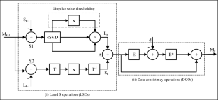
Methods: Low-rank plus sparse (L+S) matrix decomposition model is a technique used in literature to reconstruct the highly undersampled dynamic MRI (dMRI) data at the expense of long reconstruction time. In this paper, Compressed Singular Value Decomposition (cSVD) model is used in L+S decomposition model (instead of conventional SVD) to reduce the reconstruction time. The results provide improved quality of the reconstructed images. Furthermore, it has been observed that cSVD and other parts of the L+S model possess highly parallel operations; therefore, a customized GPU based parallel architecture of the modified L+S model has been presented to further reduce the reconstruction time.
Results: Four cardiac MRI datasets (three different cardiac perfusion acquired from different patients and one cardiac cine data), each with different acceleration factors of 2, 6 and 8 are used for experiments in this paper. Experimental results demonstrate that using the proposed parallel architecture for the reconstruction of cardiac perfusion data provides a speed-up factor up to 19.15× (with memory latency) and 70.55× (without memory latency) in comparison to the conventional CPU reconstruction with no compromise on image quality.
Conclusion: The proposed method is well-suited for real-time clinical applications, offering a substantial reduction in reconstruction time.
期刊介绍:
MAGMA is a multidisciplinary international journal devoted to the publication of articles on all aspects of magnetic resonance techniques and their applications in medicine and biology. MAGMA currently publishes research papers, reviews, letters to the editor, and commentaries, six times a year. The subject areas covered by MAGMA include:
advances in materials, hardware and software in magnetic resonance technology,
new developments and results in research and practical applications of magnetic resonance imaging and spectroscopy related to biology and medicine,
study of animal models and intact cells using magnetic resonance,
reports of clinical trials on humans and clinical validation of magnetic resonance protocols.

 求助内容:
求助内容: 应助结果提醒方式:


