弥散加权磁共振成像ADC值在1.5 T乳腺病变鉴别诊断中的诊断价值:系统综述和荟萃分析

IF 1.6 4区 医学 Q4 ENGINEERING, BIOMEDICAL
Winniecia Dkhar, Rajagopal Kadavigere, Suresh Sukumar, Abhimanyu Pradhan, S Sharath
{"title":"弥散加权磁共振成像ADC值在1.5 T乳腺病变鉴别诊断中的诊断价值:系统综述和荟萃分析","authors":"Winniecia Dkhar, Rajagopal Kadavigere, Suresh Sukumar, Abhimanyu Pradhan, S Sharath","doi":"10.1007/s40846-023-00825-9","DOIUrl":null,"url":null,"abstract":"Abstract Purpose Medical technology has gone a long way in diagnosis and characterization of breast tumors. Diffusion-weighted MR imaging is the state of the art for breast screening and diagnosing. The aim of this meta-analysis is to evaluate the diagnostic performances of diffusion-weighted MR imaging in characterization of breast lesions with different b value in 1.5 T MRI. Method An extensive search on Scopus, Embase, and PubMed databases were performed on studies published between January 2000 and 2020. The systematic seek initially yielded 2467 studies, out of which 27 research were covered on this meta-evaluation. The included studies for meta-analysis utilized different b value and noted that the ADC value was highly influenced by the b value, for differential diagnosis of breast tumors. Results The current meta-analysis has shown the ADC values was lower for malignant breast lesions as compared with benign lesions. The recommended mean threshold ADC was 1.25 ± 0.17 × 10 –3 mm 2 /s range from 0.93 to 1.60 × 10 –3 mm 2 /s for differential diagnosis of breast tumors. Sub-group analysis on the bases of b value showed statistically significant differences in the ADC value of benign and malignant breast tumors. Conclusion In conclusion, we noted that b value has a significant effect in calculating the ADC value of the breast lesions as well as ADC threshold value but lacks standardization. The ADC value measurement has a potential for differentiation between benign and malignant breast lesions.","PeriodicalId":50133,"journal":{"name":"Journal of Medical and Biological Engineering","volume":null,"pages":null},"PeriodicalIF":1.6000,"publicationDate":"2023-09-21","publicationTypes":"Journal Article","fieldsOfStudy":null,"isOpenAccess":false,"openAccessPdf":"","citationCount":"0","resultStr":"{\"title\":\"Diagnostic Performances of ADC Value in Diffusion-Weighted MR Imaging for Differential Diagnosis of Breast Lesions in 1.5 T: A Systematic Review and Meta-analysis\",\"authors\":\"Winniecia Dkhar, Rajagopal Kadavigere, Suresh Sukumar, Abhimanyu Pradhan, S Sharath\",\"doi\":\"10.1007/s40846-023-00825-9\",\"DOIUrl\":null,\"url\":null,\"abstract\":\"Abstract Purpose Medical technology has gone a long way in diagnosis and characterization of breast tumors. Diffusion-weighted MR imaging is the state of the art for breast screening and diagnosing. The aim of this meta-analysis is to evaluate the diagnostic performances of diffusion-weighted MR imaging in characterization of breast lesions with different b value in 1.5 T MRI. Method An extensive search on Scopus, Embase, and PubMed databases were performed on studies published between January 2000 and 2020. The systematic seek initially yielded 2467 studies, out of which 27 research were covered on this meta-evaluation. The included studies for meta-analysis utilized different b value and noted that the ADC value was highly influenced by the b value, for differential diagnosis of breast tumors. Results The current meta-analysis has shown the ADC values was lower for malignant breast lesions as compared with benign lesions. The recommended mean threshold ADC was 1.25 ± 0.17 × 10 –3 mm 2 /s range from 0.93 to 1.60 × 10 –3 mm 2 /s for differential diagnosis of breast tumors. Sub-group analysis on the bases of b value showed statistically significant differences in the ADC value of benign and malignant breast tumors. Conclusion In conclusion, we noted that b value has a significant effect in calculating the ADC value of the breast lesions as well as ADC threshold value but lacks standardization. The ADC value measurement has a potential for differentiation between benign and malignant breast lesions.\",\"PeriodicalId\":50133,\"journal\":{\"name\":\"Journal of Medical and Biological Engineering\",\"volume\":null,\"pages\":null},\"PeriodicalIF\":1.6000,\"publicationDate\":\"2023-09-21\",\"publicationTypes\":\"Journal Article\",\"fieldsOfStudy\":null,\"isOpenAccess\":false,\"openAccessPdf\":\"\",\"citationCount\":\"0\",\"resultStr\":null,\"platform\":\"Semanticscholar\",\"paperid\":null,\"PeriodicalName\":\"Journal of Medical and Biological Engineering\",\"FirstCategoryId\":\"1085\",\"ListUrlMain\":\"https://doi.org/10.1007/s40846-023-00825-9\",\"RegionNum\":4,\"RegionCategory\":\"医学\",\"ArticlePicture\":[],\"TitleCN\":null,\"AbstractTextCN\":null,\"PMCID\":null,\"EPubDate\":\"\",\"PubModel\":\"\",\"JCR\":\"Q4\",\"JCRName\":\"ENGINEERING, BIOMEDICAL\",\"Score\":null,\"Total\":0}","platform":"Semanticscholar","paperid":null,"PeriodicalName":"Journal of Medical and Biological Engineering","FirstCategoryId":"1085","ListUrlMain":"https://doi.org/10.1007/s40846-023-00825-9","RegionNum":4,"RegionCategory":"医学","ArticlePicture":[],"TitleCN":null,"AbstractTextCN":null,"PMCID":null,"EPubDate":"","PubModel":"","JCR":"Q4","JCRName":"ENGINEERING, BIOMEDICAL","Score":null,"Total":0}
引用次数: 0

摘要

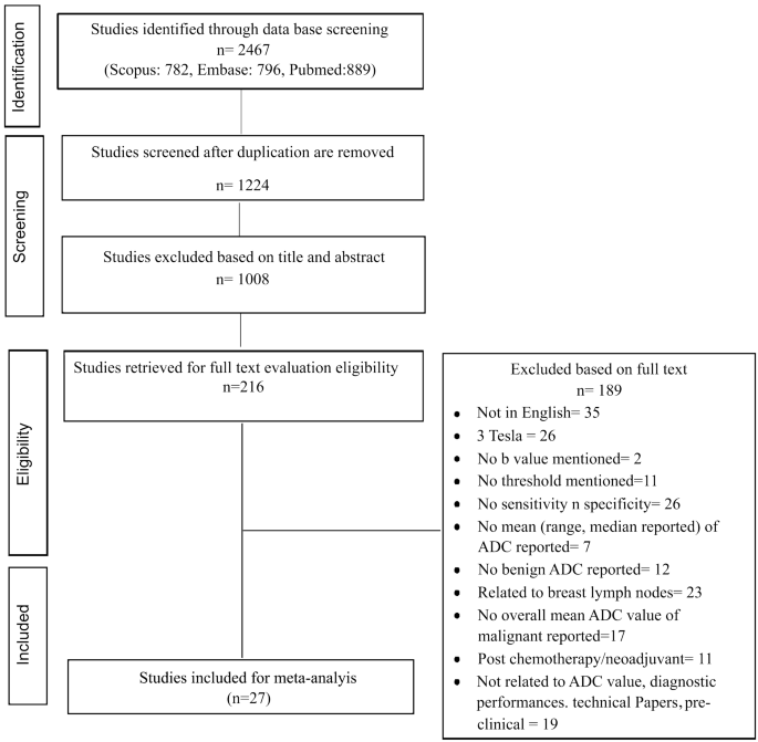
摘要目的医学技术在乳腺肿瘤的诊断和表征方面取得了长足的进步。弥散加权磁共振成像是乳腺筛查和诊断的最新技术。本荟萃分析的目的是评估扩散加权磁共振成像在1.5 T MRI不同b值乳腺病变特征中的诊断性能。方法广泛检索2000年1月至2020年1月间发表的Scopus、Embase和PubMed数据库。系统搜索最初产生了2467项研究,其中27项研究被纳入本meta评价。纳入meta分析的研究使用了不同的b值,并注意到ADC值受b值的高度影响,用于乳腺肿瘤的鉴别诊断。结果目前的荟萃分析显示,乳腺恶性病变的ADC值低于良性病变。乳腺肿瘤鉴别诊断推荐的平均阈值ADC为1.25±0.17 × 10 - 3mm2 /s,范围为0.93 ~ 1.60 × 10 - 3mm2 /s。以b值为基础进行亚组分析,良恶性乳腺肿瘤的ADC值差异有统计学意义。综上所述,我们注意到b值在计算乳腺病变ADC值和ADC阈值方面有显著作用,但缺乏标准化。ADC值测量具有区分乳腺良恶性病变的潜力。
本文章由计算机程序翻译,如有差异,请以英文原文为准。

Diagnostic Performances of ADC Value in Diffusion-Weighted MR Imaging for Differential Diagnosis of Breast Lesions in 1.5 T: A Systematic Review and Meta-analysis

Diagnostic Performances of ADC Value in Diffusion-Weighted MR Imaging for Differential Diagnosis of Breast Lesions in 1.5 T: A Systematic Review and Meta-analysis
Abstract Purpose Medical technology has gone a long way in diagnosis and characterization of breast tumors. Diffusion-weighted MR imaging is the state of the art for breast screening and diagnosing. The aim of this meta-analysis is to evaluate the diagnostic performances of diffusion-weighted MR imaging in characterization of breast lesions with different b value in 1.5 T MRI. Method An extensive search on Scopus, Embase, and PubMed databases were performed on studies published between January 2000 and 2020. The systematic seek initially yielded 2467 studies, out of which 27 research were covered on this meta-evaluation. The included studies for meta-analysis utilized different b value and noted that the ADC value was highly influenced by the b value, for differential diagnosis of breast tumors. Results The current meta-analysis has shown the ADC values was lower for malignant breast lesions as compared with benign lesions. The recommended mean threshold ADC was 1.25 ± 0.17 × 10 –3 mm 2 /s range from 0.93 to 1.60 × 10 –3 mm 2 /s for differential diagnosis of breast tumors. Sub-group analysis on the bases of b value showed statistically significant differences in the ADC value of benign and malignant breast tumors. Conclusion In conclusion, we noted that b value has a significant effect in calculating the ADC value of the breast lesions as well as ADC threshold value but lacks standardization. The ADC value measurement has a potential for differentiation between benign and malignant breast lesions.
求助全文
通过发布文献求助,成功后即可免费获取论文全文。 去求助
来源期刊
CiteScore
4.30
自引率
5.00%
发文量
81
审稿时长
3 months
期刊介绍: The purpose of Journal of Medical and Biological Engineering, JMBE, is committed to encouraging and providing the standard of biomedical engineering. The journal is devoted to publishing papers related to clinical engineering, biomedical signals, medical imaging, bio-informatics, tissue engineering, and so on. Other than the above articles, any contributions regarding hot issues and technological developments that help reach the purpose are also included.
×
引用
GB/T 7714-2015
复制
MLA
复制
APA
复制
导出至
BibTeX EndNote RefMan NoteFirst NoteExpress
×
提示
您的信息不完整,为了账户安全,请先补充。
现在去补充
×
提示
您因"违规操作"
具体请查看互助需知
我知道了
×
提示
确定
请完成安全验证×
copy
已复制链接
快去分享给好友吧!
我知道了
右上角分享
点击右上角分享
0
联系我们:info@booksci.cn Book学术提供免费学术资源搜索服务,方便国内外学者检索中英文文献。致力于提供最便捷和优质的服务体验。 Copyright © 2023 布克学术 All rights reserved.
京ICP备2023020795号-1
ghs 京公网安备 11010802042870号
Book学术文献互助
Book学术文献互助群
群 号:481959085
Book学术官方微信