Juanwei Ma , Kaizhong Xue , Xinyu Wang , Mengjing Cai , Xinli Wang , Jiaojiao Li , Linlin Song , He Wang , Yali Niu , Jing Wang , Zhaoxiang Ye , Jing Zhang , Feng Liu
{"title":"血管性认知障碍的灰质体积异常及其与基因表达谱的关系","authors":"Juanwei Ma , Kaizhong Xue , Xinyu Wang , Mengjing Cai , Xinli Wang , Jiaojiao Li , Linlin Song , He Wang , Yali Niu , Jing Wang , Zhaoxiang Ye , Jing Zhang , Feng Liu","doi":"10.1016/j.metrad.2023.100035","DOIUrl":null,"url":null,"abstract":"<div><h3>Background</h3><p>It has been revealed that brain gray matter volume (GMV) abnormalities are present in patients with vascular cognitive impairment (VCI). However, the GMV alterations that have been uncovered are highly inconsistent, and their correlation with gene expression profiles is still largely unknown.</p></div><div><h3>Purpose</h3><p>To establish a correlation between VCI-related GMV alterations and gene expression patterns and uncover potential genetic profiles underlying GMV abnormalities in VCI.</p></div><div><h3>Materials and methods</h3><p>Here, a quantitative meta-analysis that compared voxel-based GMV between VCI patients and healthy controls (HCs) was carried out on 11 datasets (10 from previous studies and 1 newly collected), comprising 385 VCI individuals and 334 HCs, to investigate GMV alterations in VCI patients. Partial least squares regression analysis was then conducted to investigate the relationship between the GMV alterations in VCI and gene expression profiles obtained from Allen Human Brain Atlas database.</p></div><div><h3>Results</h3><p>Compared with healthy controls, patients with VCI showed consistent decreased GMV which predominantly included the right insula, right Rolandic operculum, right putamen, right superior temporal gyrus, left medial superior frontal gyrus, and right median cingulate and paracingulate gyri. Meta-regression analysis revealed that decreased GMV in left medial superior frontal gyrus was negatively correlated with Mini-Mental State Examination score in VCI. Furthermore, 2835 genes were identified whose expression patterns were correlated with VCI-related GMV changes, and these genes were enriched in distinct biological processes, brain cell types and lifespan windows across brain regions.</p></div><div><h3>Conclusion</h3><p>Together, these findings could provide the potential neurobiological underpinnings and the genetic substrates underlying GMV abnormalities of VCI.</p></div>","PeriodicalId":100921,"journal":{"name":"Meta-Radiology","volume":"1 3","pages":"Article 100035"},"PeriodicalIF":0.0000,"publicationDate":"2023-11-01","publicationTypes":"Journal Article","fieldsOfStudy":null,"isOpenAccess":false,"openAccessPdf":"https://www.sciencedirect.com/science/article/pii/S2950162823000358/pdfft?md5=a02cc6787e2fadef10646a1cbbec8e75&pid=1-s2.0-S2950162823000358-main.pdf","citationCount":"0","resultStr":"{\"title\":\"Gray matter volume abnormalities in vascular cognitive impairment and their association with gene expression profiles\",\"authors\":\"Juanwei Ma , Kaizhong Xue , Xinyu Wang , Mengjing Cai , Xinli Wang , Jiaojiao Li , Linlin Song , He Wang , Yali Niu , Jing Wang , Zhaoxiang Ye , Jing Zhang , Feng Liu\",\"doi\":\"10.1016/j.metrad.2023.100035\",\"DOIUrl\":null,\"url\":null,\"abstract\":\"<div><h3>Background</h3><p>It has been revealed that brain gray matter volume (GMV) abnormalities are present in patients with vascular cognitive impairment (VCI). However, the GMV alterations that have been uncovered are highly inconsistent, and their correlation with gene expression profiles is still largely unknown.</p></div><div><h3>Purpose</h3><p>To establish a correlation between VCI-related GMV alterations and gene expression patterns and uncover potential genetic profiles underlying GMV abnormalities in VCI.</p></div><div><h3>Materials and methods</h3><p>Here, a quantitative meta-analysis that compared voxel-based GMV between VCI patients and healthy controls (HCs) was carried out on 11 datasets (10 from previous studies and 1 newly collected), comprising 385 VCI individuals and 334 HCs, to investigate GMV alterations in VCI patients. Partial least squares regression analysis was then conducted to investigate the relationship between the GMV alterations in VCI and gene expression profiles obtained from Allen Human Brain Atlas database.</p></div><div><h3>Results</h3><p>Compared with healthy controls, patients with VCI showed consistent decreased GMV which predominantly included the right insula, right Rolandic operculum, right putamen, right superior temporal gyrus, left medial superior frontal gyrus, and right median cingulate and paracingulate gyri. Meta-regression analysis revealed that decreased GMV in left medial superior frontal gyrus was negatively correlated with Mini-Mental State Examination score in VCI. Furthermore, 2835 genes were identified whose expression patterns were correlated with VCI-related GMV changes, and these genes were enriched in distinct biological processes, brain cell types and lifespan windows across brain regions.</p></div><div><h3>Conclusion</h3><p>Together, these findings could provide the potential neurobiological underpinnings and the genetic substrates underlying GMV abnormalities of VCI.</p></div>\",\"PeriodicalId\":100921,\"journal\":{\"name\":\"Meta-Radiology\",\"volume\":\"1 3\",\"pages\":\"Article 100035\"},\"PeriodicalIF\":0.0000,\"publicationDate\":\"2023-11-01\",\"publicationTypes\":\"Journal Article\",\"fieldsOfStudy\":null,\"isOpenAccess\":false,\"openAccessPdf\":\"https://www.sciencedirect.com/science/article/pii/S2950162823000358/pdfft?md5=a02cc6787e2fadef10646a1cbbec8e75&pid=1-s2.0-S2950162823000358-main.pdf\",\"citationCount\":\"0\",\"resultStr\":null,\"platform\":\"Semanticscholar\",\"paperid\":null,\"PeriodicalName\":\"Meta-Radiology\",\"FirstCategoryId\":\"1085\",\"ListUrlMain\":\"https://www.sciencedirect.com/science/article/pii/S2950162823000358\",\"RegionNum\":0,\"RegionCategory\":null,\"ArticlePicture\":[],\"TitleCN\":null,\"AbstractTextCN\":null,\"PMCID\":null,\"EPubDate\":\"\",\"PubModel\":\"\",\"JCR\":\"\",\"JCRName\":\"\",\"Score\":null,\"Total\":0}","platform":"Semanticscholar","paperid":null,"PeriodicalName":"Meta-Radiology","FirstCategoryId":"1085","ListUrlMain":"https://www.sciencedirect.com/science/article/pii/S2950162823000358","RegionNum":0,"RegionCategory":null,"ArticlePicture":[],"TitleCN":null,"AbstractTextCN":null,"PMCID":null,"EPubDate":"","PubModel":"","JCR":"","JCRName":"","Score":null,"Total":0}
引用次数: 0
摘要
研究发现血管性认知障碍(VCI)患者存在脑灰质体积(GMV)异常。然而,已经发现的GMV改变是高度不一致的,它们与基因表达谱的相关性仍然很大程度上是未知的。建立VCI相关GMV改变与基因表达模式之间的相关性,揭示VCI中GMV异常的潜在遗传谱。本研究对11个数据集(10个来自先前的研究,1个新收集的数据集)进行了定量荟萃分析,比较了VCI患者和健康对照(hc)之间基于体素的GMV,包括385个VCI个体和334个hc,以调查VCI患者的GMV变化。通过偏最小二乘回归分析,探讨VCI GMV变化与Allen Human Brain Atlas数据库中基因表达谱的关系。与健康对照组相比,VCI患者GMV持续下降,主要包括右脑岛、右罗兰底盖、右壳核、右颞上回、左额叶内侧上回、右扣带和副扣带中回。meta回归分析显示,左内侧额上回GMV下降与VCI小精神状态检查得分呈负相关。此外,还鉴定出2835个基因的表达模式与vci相关的GMV变化相关,这些基因在不同的生物过程、脑细胞类型和大脑区域的寿命窗口中富集。总之,这些发现可能为VCI的GMV异常提供潜在的神经生物学基础和遗传基础。
Gray matter volume abnormalities in vascular cognitive impairment and their association with gene expression profiles
Background
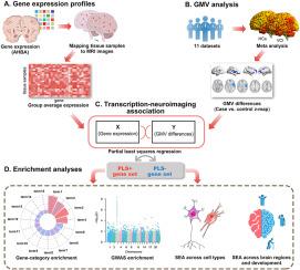
It has been revealed that brain gray matter volume (GMV) abnormalities are present in patients with vascular cognitive impairment (VCI). However, the GMV alterations that have been uncovered are highly inconsistent, and their correlation with gene expression profiles is still largely unknown.
Purpose
To establish a correlation between VCI-related GMV alterations and gene expression patterns and uncover potential genetic profiles underlying GMV abnormalities in VCI.
Materials and methods
Here, a quantitative meta-analysis that compared voxel-based GMV between VCI patients and healthy controls (HCs) was carried out on 11 datasets (10 from previous studies and 1 newly collected), comprising 385 VCI individuals and 334 HCs, to investigate GMV alterations in VCI patients. Partial least squares regression analysis was then conducted to investigate the relationship between the GMV alterations in VCI and gene expression profiles obtained from Allen Human Brain Atlas database.
Results
Compared with healthy controls, patients with VCI showed consistent decreased GMV which predominantly included the right insula, right Rolandic operculum, right putamen, right superior temporal gyrus, left medial superior frontal gyrus, and right median cingulate and paracingulate gyri. Meta-regression analysis revealed that decreased GMV in left medial superior frontal gyrus was negatively correlated with Mini-Mental State Examination score in VCI. Furthermore, 2835 genes were identified whose expression patterns were correlated with VCI-related GMV changes, and these genes were enriched in distinct biological processes, brain cell types and lifespan windows across brain regions.
Conclusion
Together, these findings could provide the potential neurobiological underpinnings and the genetic substrates underlying GMV abnormalities of VCI.

 求助内容:
求助内容: 应助结果提醒方式:


