Emiliya Usheva, Dustin Williams, Haley Musgrave, Scott Zhou
{"title":"超声球后斑点征象诊断视网膜中央动脉闭塞1例。","authors":"Emiliya Usheva, Dustin Williams, Haley Musgrave, Scott Zhou","doi":"10.21980/J8735P","DOIUrl":null,"url":null,"abstract":"<p><p>Central retinal artery occlusion (CRAO) is a rare emergency department presentation with high morbidity and potential for long-term vision loss. Additionally, this finding requires an expeditious embolic workup for possible systemic pathology (i.e., stroke). The gold standard for diagnosis is visualization of a pale retina with a \"cherry-red spot\" on the fovea seen under dilated fundoscopic examination. However, performing a dilated fundoscopic exam is often not practical and technically challenging in the emergency room setting. Alternatively, point of care ultrasound is an inexpensive, non-invasive tool that is already highly utilized in the emergency department and can aid in diagnosis. In the case described in this report, a 66-year-old female presented to the emergency department with painless, monocular vision loss. Ultrasound showed a hyperechoic density on the distal aspect of the optic nerve (\"retrobulbar spot sign\") and dilated fundoscopic exam showed right eye pale macula with cherry red spot, all consistent with CRAO. Here we present a case that suggests an opportunity for improvement in evaluation of monocular vision loss in the emergency department by adding bedside ocular ultrasound to aid in more rapid diagnosis of CRAO.</p><p><strong>Topics: </strong>Central retinal occlusion, vision loss, point-of-care ultrasound, ocular ultrasound, emboli.</p>","PeriodicalId":73721,"journal":{"name":"Journal of education & teaching in emergency medicine","volume":"8 4","pages":"V5-V8"},"PeriodicalIF":0.0000,"publicationDate":"2023-10-31","publicationTypes":"Journal Article","fieldsOfStudy":null,"isOpenAccess":false,"openAccessPdf":"https://www.ncbi.nlm.nih.gov/pmc/articles/PMC10631812/pdf/","citationCount":"0","resultStr":"{\"title\":\"Sonographic Retrobulbar Spot Sign in Diagnosis of Central Retinal Artery Occlusion: A Case Report.\",\"authors\":\"Emiliya Usheva, Dustin Williams, Haley Musgrave, Scott Zhou\",\"doi\":\"10.21980/J8735P\",\"DOIUrl\":null,\"url\":null,\"abstract\":\"<p><p>Central retinal artery occlusion (CRAO) is a rare emergency department presentation with high morbidity and potential for long-term vision loss. Additionally, this finding requires an expeditious embolic workup for possible systemic pathology (i.e., stroke). The gold standard for diagnosis is visualization of a pale retina with a \\\"cherry-red spot\\\" on the fovea seen under dilated fundoscopic examination. However, performing a dilated fundoscopic exam is often not practical and technically challenging in the emergency room setting. Alternatively, point of care ultrasound is an inexpensive, non-invasive tool that is already highly utilized in the emergency department and can aid in diagnosis. In the case described in this report, a 66-year-old female presented to the emergency department with painless, monocular vision loss. Ultrasound showed a hyperechoic density on the distal aspect of the optic nerve (\\\"retrobulbar spot sign\\\") and dilated fundoscopic exam showed right eye pale macula with cherry red spot, all consistent with CRAO. Here we present a case that suggests an opportunity for improvement in evaluation of monocular vision loss in the emergency department by adding bedside ocular ultrasound to aid in more rapid diagnosis of CRAO.</p><p><strong>Topics: </strong>Central retinal occlusion, vision loss, point-of-care ultrasound, ocular ultrasound, emboli.</p>\",\"PeriodicalId\":73721,\"journal\":{\"name\":\"Journal of education & teaching in emergency medicine\",\"volume\":\"8 4\",\"pages\":\"V5-V8\"},\"PeriodicalIF\":0.0000,\"publicationDate\":\"2023-10-31\",\"publicationTypes\":\"Journal Article\",\"fieldsOfStudy\":null,\"isOpenAccess\":false,\"openAccessPdf\":\"https://www.ncbi.nlm.nih.gov/pmc/articles/PMC10631812/pdf/\",\"citationCount\":\"0\",\"resultStr\":null,\"platform\":\"Semanticscholar\",\"paperid\":null,\"PeriodicalName\":\"Journal of education & teaching in emergency medicine\",\"FirstCategoryId\":\"1085\",\"ListUrlMain\":\"https://doi.org/10.21980/J8735P\",\"RegionNum\":0,\"RegionCategory\":null,\"ArticlePicture\":[],\"TitleCN\":null,\"AbstractTextCN\":null,\"PMCID\":null,\"EPubDate\":\"2023/10/1 0:00:00\",\"PubModel\":\"eCollection\",\"JCR\":\"\",\"JCRName\":\"\",\"Score\":null,\"Total\":0}","platform":"Semanticscholar","paperid":null,"PeriodicalName":"Journal of education & teaching in emergency medicine","FirstCategoryId":"1085","ListUrlMain":"https://doi.org/10.21980/J8735P","RegionNum":0,"RegionCategory":null,"ArticlePicture":[],"TitleCN":null,"AbstractTextCN":null,"PMCID":null,"EPubDate":"2023/10/1 0:00:00","PubModel":"eCollection","JCR":"","JCRName":"","Score":null,"Total":0}
Sonographic Retrobulbar Spot Sign in Diagnosis of Central Retinal Artery Occlusion: A Case Report.
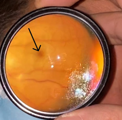
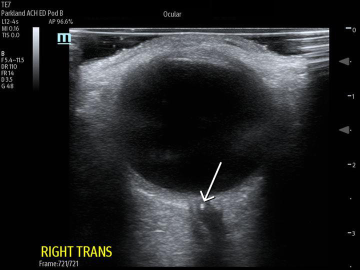
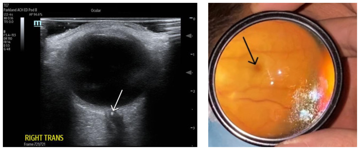
Central retinal artery occlusion (CRAO) is a rare emergency department presentation with high morbidity and potential for long-term vision loss. Additionally, this finding requires an expeditious embolic workup for possible systemic pathology (i.e., stroke). The gold standard for diagnosis is visualization of a pale retina with a "cherry-red spot" on the fovea seen under dilated fundoscopic examination. However, performing a dilated fundoscopic exam is often not practical and technically challenging in the emergency room setting. Alternatively, point of care ultrasound is an inexpensive, non-invasive tool that is already highly utilized in the emergency department and can aid in diagnosis. In the case described in this report, a 66-year-old female presented to the emergency department with painless, monocular vision loss. Ultrasound showed a hyperechoic density on the distal aspect of the optic nerve ("retrobulbar spot sign") and dilated fundoscopic exam showed right eye pale macula with cherry red spot, all consistent with CRAO. Here we present a case that suggests an opportunity for improvement in evaluation of monocular vision loss in the emergency department by adding bedside ocular ultrasound to aid in more rapid diagnosis of CRAO.

 求助内容:
求助内容: 应助结果提醒方式:


