根据活检部位不同组织学表现诊断新发麻风患者。

IF 1.5 4区 医学 Q3 DERMATOLOGY
Shinyoung Song, Jaewoo Lim, Seha Park, Seulki Lee, Jinok Baek, Hyangjoon Park, Joo Young Roh, Hee Joo Kim
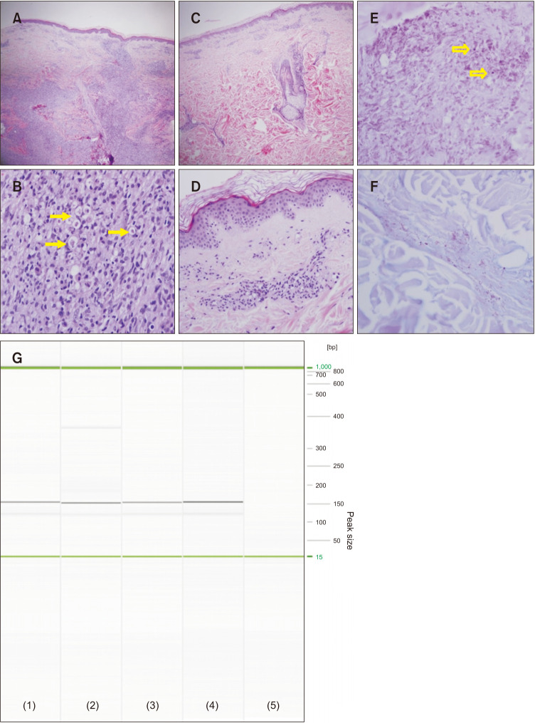
{"title":"根据活检部位不同组织学表现诊断新发麻风患者。","authors":"Shinyoung Song, Jaewoo Lim, Seha Park, Seulki Lee, Jinok Baek, Hyangjoon Park, Joo Young Roh, Hee Joo Kim","doi":"10.5021/ad.20.224","DOIUrl":null,"url":null,"abstract":": An 81-year-old male had multiple erythematous to dusky patches on the trunk, arms, and legs for several years (Fig. 1A, B) and two violaceous nodules on the back with unknown onset (Fig. 1C). He had numbness of the hands and feet and edema of the hands and face during the previous six weeks. Punch biopsies were performed on the patch and nodule on the back. The nodule showed dense dermal infiltration by foamy histiocytes and lepra cells containing degenerated microbial components (Fig. 2A, B). The patch showed only superficial dermal perivascular and periappendageal lymphohistiocytic infiltration (Fig. 2C, D). Acid-fast bacillus (AFB) staining revealed that acid-fast bacilli was denser in the nodule than in the patch and some of the bacilli were clustered, forming globi (Fig. 2E, F). The patient was diagnosed as a new lepromatous leprosy patient based on clinical and histological findings. The patient was referred to the Hansen Welfare Association of Korea. Amplication of 154 bp Mycobacterium leprae-specific repetitive element (RLEP) was confirmed in PCR results of Hansen Welfare Association of Korea (Fig. 2G). The institution confirmed that the patient was","PeriodicalId":8233,"journal":{"name":"Annals of Dermatology","volume":"34 6","pages":"487-489"},"PeriodicalIF":1.5000,"publicationDate":"2022-12-01","publicationTypes":"Journal Article","fieldsOfStudy":null,"isOpenAccess":false,"openAccessPdf":"https://ftp.ncbi.nlm.nih.gov/pub/pmc/oa_pdf/0e/b6/ad-34-487.PMC9763914.pdf","citationCount":"0","resultStr":"{\"title\":\"Diagnosis of New Leprosy Patients through Various Histological Findings according to Biopsy Sites.\",\"authors\":\"Shinyoung Song, Jaewoo Lim, Seha Park, Seulki Lee, Jinok Baek, Hyangjoon Park, Joo Young Roh, Hee Joo Kim\",\"doi\":\"10.5021/ad.20.224\",\"DOIUrl\":null,\"url\":null,\"abstract\":\": An 81-year-old male had multiple erythematous to dusky patches on the trunk, arms, and legs for several years (Fig. 1A, B) and two violaceous nodules on the back with unknown onset (Fig. 1C). He had numbness of the hands and feet and edema of the hands and face during the previous six weeks. Punch biopsies were performed on the patch and nodule on the back. The nodule showed dense dermal infiltration by foamy histiocytes and lepra cells containing degenerated microbial components (Fig. 2A, B). The patch showed only superficial dermal perivascular and periappendageal lymphohistiocytic infiltration (Fig. 2C, D). Acid-fast bacillus (AFB) staining revealed that acid-fast bacilli was denser in the nodule than in the patch and some of the bacilli were clustered, forming globi (Fig. 2E, F). The patient was diagnosed as a new lepromatous leprosy patient based on clinical and histological findings. The patient was referred to the Hansen Welfare Association of Korea. Amplication of 154 bp Mycobacterium leprae-specific repetitive element (RLEP) was confirmed in PCR results of Hansen Welfare Association of Korea (Fig. 2G). The institution confirmed that the patient was\",\"PeriodicalId\":8233,\"journal\":{\"name\":\"Annals of Dermatology\",\"volume\":\"34 6\",\"pages\":\"487-489\"},\"PeriodicalIF\":1.5000,\"publicationDate\":\"2022-12-01\",\"publicationTypes\":\"Journal Article\",\"fieldsOfStudy\":null,\"isOpenAccess\":false,\"openAccessPdf\":\"https://ftp.ncbi.nlm.nih.gov/pub/pmc/oa_pdf/0e/b6/ad-34-487.PMC9763914.pdf\",\"citationCount\":\"0\",\"resultStr\":null,\"platform\":\"Semanticscholar\",\"paperid\":null,\"PeriodicalName\":\"Annals of Dermatology\",\"FirstCategoryId\":\"3\",\"ListUrlMain\":\"https://doi.org/10.5021/ad.20.224\",\"RegionNum\":4,\"RegionCategory\":\"医学\",\"ArticlePicture\":[],\"TitleCN\":null,\"AbstractTextCN\":null,\"PMCID\":null,\"EPubDate\":\"\",\"PubModel\":\"\",\"JCR\":\"Q3\",\"JCRName\":\"DERMATOLOGY\",\"Score\":null,\"Total\":0}","platform":"Semanticscholar","paperid":null,"PeriodicalName":"Annals of Dermatology","FirstCategoryId":"3","ListUrlMain":"https://doi.org/10.5021/ad.20.224","RegionNum":4,"RegionCategory":"医学","ArticlePicture":[],"TitleCN":null,"AbstractTextCN":null,"PMCID":null,"EPubDate":"","PubModel":"","JCR":"Q3","JCRName":"DERMATOLOGY","Score":null,"Total":0}
引用次数: 0

摘要

本文章由计算机程序翻译,如有差异,请以英文原文为准。
Diagnosis of New Leprosy Patients through Various Histological Findings according to Biopsy Sites.

Diagnosis of New Leprosy Patients through Various Histological Findings according to Biopsy Sites.

Diagnosis of New Leprosy Patients through Various Histological Findings according to Biopsy Sites.
: An 81-year-old male had multiple erythematous to dusky patches on the trunk, arms, and legs for several years (Fig. 1A, B) and two violaceous nodules on the back with unknown onset (Fig. 1C). He had numbness of the hands and feet and edema of the hands and face during the previous six weeks. Punch biopsies were performed on the patch and nodule on the back. The nodule showed dense dermal infiltration by foamy histiocytes and lepra cells containing degenerated microbial components (Fig. 2A, B). The patch showed only superficial dermal perivascular and periappendageal lymphohistiocytic infiltration (Fig. 2C, D). Acid-fast bacillus (AFB) staining revealed that acid-fast bacilli was denser in the nodule than in the patch and some of the bacilli were clustered, forming globi (Fig. 2E, F). The patient was diagnosed as a new lepromatous leprosy patient based on clinical and histological findings. The patient was referred to the Hansen Welfare Association of Korea. Amplication of 154 bp Mycobacterium leprae-specific repetitive element (RLEP) was confirmed in PCR results of Hansen Welfare Association of Korea (Fig. 2G). The institution confirmed that the patient was
求助全文
通过发布文献求助,成功后即可免费获取论文全文。 去求助
来源期刊
Annals of Dermatology
Annals of Dermatology 医学-皮肤病学
CiteScore
1.60
自引率
6.20%
发文量
77
审稿时长
6-12 weeks
期刊介绍: Annals of Dermatology (Ann Dermatol) is the official peer-reviewed publication of the Korean Dermatological Association and the Korean Society for Investigative Dermatology. Since 1989, Ann Dermatol has contributed as a platform for communicating the latest research outcome and recent trend of dermatology in Korea and all over the world. Ann Dermatol seeks for ameliorated understanding of skin and skin-related disease for clinicians and researchers. Ann Dermatol deals with diverse skin-related topics from laboratory investigations to clinical outcomes and invites review articles, original articles, case reports, brief reports and items of correspondence. Ann Dermatol is interested in contributions from all countries in which good and advanced research is carried out. Ann Dermatol willingly recruits well-organized and significant manuscripts with proper scope throughout the world.
×
引用
GB/T 7714-2015
复制
MLA
复制
APA
复制
导出至
BibTeX EndNote RefMan NoteFirst NoteExpress
×
提示
您的信息不完整,为了账户安全,请先补充。
现在去补充
×
提示
您因"违规操作"
具体请查看互助需知
我知道了
×
提示
确定
请完成安全验证×
copy
已复制链接
快去分享给好友吧!
我知道了
右上角分享
点击右上角分享
0
联系我们:info@booksci.cn Book学术提供免费学术资源搜索服务,方便国内外学者检索中英文文献。致力于提供最便捷和优质的服务体验。 Copyright © 2023 布克学术 All rights reserved.
京ICP备2023020795号-1
ghs 京公网安备 11010802042870号
Book学术文献互助
Book学术文献互助群
群 号:481959085
Book学术官方微信