Janaka Senarathna, Mark Kovler, Ayush Prasad, Akanksha Bhargava, Nitish V. Thakor, Chhinder P. Sodhi, David J. Hackam, Arvind P. Pathak
{"title":"多对比光学成像研究坏死性小肠结肠炎微血管的体内表型","authors":"Janaka Senarathna, Mark Kovler, Ayush Prasad, Akanksha Bhargava, Nitish V. Thakor, Chhinder P. Sodhi, David J. Hackam, Arvind P. Pathak","doi":"10.1111/micc.12768","DOIUrl":null,"url":null,"abstract":"<div>\n \n \n <section>\n \n <h3> Objective</h3>\n \n <p>Necrotizing enterocolitis (NEC) is the most prevalent gastrointestinal emergency in premature infants and is characterized by a dysfunctional gut microcirculation. Therefore, there is a dire need for in vivo methods to characterize NEC-induced changes in the structure and function of the gut microcirculation, that is, its vascular phenotype. Since in vivo gut imaging methods are often slow and employ a single-contrast mechanism, we developed a rapid multicontrast imaging technique and a novel analyses pipeline for phenotyping the gut microcirculation.</p>\n </section>\n \n <section>\n \n <h3> Methods</h3>\n \n <p>Using an experimental NEC model, we acquired in vivo images of the gut microvasculature and blood flow over a 5000 × 7000 μm<sup>2</sup> field of view at 5 μm resolution via the following two endogenous contrast mechanisms: intrinsic optical signals and laser speckles. Next, we transformed intestinal images into rectilinear “flat maps,” and delineated 1A/V gut microvessels and their perfusion territories as “intestinal vascular units” (IVUs). Employing IVUs, we quantified and visualized NEC-induced changes to the gut vascular phenotype.</p>\n </section>\n \n <section>\n \n <h3> Results</h3>\n \n <p>In vivo imaging required 60–100 s per animal. Relative to the healthy gut, NEC intestines showed a significant overall decrease (i.e. 64–72%) in perfusion, accompanied by vasoconstriction (i.e. 9–12%) and a reduction in perfusion entropy (19%)within sections of the vascular bed.</p>\n </section>\n \n <section>\n \n <h3> Conclusions</h3>\n \n <p>Multicontrast imaging coupled with IVU-based in vivo vascular phenotyping is a powerful new tool for elucidating NEC pathogenesis.</p>\n </section>\n </div>","PeriodicalId":18459,"journal":{"name":"Microcirculation","volume":null,"pages":null},"PeriodicalIF":1.9000,"publicationDate":"2022-05-20","publicationTypes":"Journal Article","fieldsOfStudy":null,"isOpenAccess":false,"openAccessPdf":"https://ftp.ncbi.nlm.nih.gov/pub/pmc/oa_pdf/2f/bb/MICC-29-e12768.PMC9633336.pdf","citationCount":"2","resultStr":"{\"title\":\"In vivo phenotyping of the microvasculature in necrotizing enterocolitis with multicontrast optical imaging\",\"authors\":\"Janaka Senarathna, Mark Kovler, Ayush Prasad, Akanksha Bhargava, Nitish V. Thakor, Chhinder P. Sodhi, David J. Hackam, Arvind P. Pathak\",\"doi\":\"10.1111/micc.12768\",\"DOIUrl\":null,\"url\":null,\"abstract\":\"<div>\\n \\n \\n <section>\\n \\n <h3> Objective</h3>\\n \\n <p>Necrotizing enterocolitis (NEC) is the most prevalent gastrointestinal emergency in premature infants and is characterized by a dysfunctional gut microcirculation. Therefore, there is a dire need for in vivo methods to characterize NEC-induced changes in the structure and function of the gut microcirculation, that is, its vascular phenotype. Since in vivo gut imaging methods are often slow and employ a single-contrast mechanism, we developed a rapid multicontrast imaging technique and a novel analyses pipeline for phenotyping the gut microcirculation.</p>\\n </section>\\n \\n <section>\\n \\n <h3> Methods</h3>\\n \\n <p>Using an experimental NEC model, we acquired in vivo images of the gut microvasculature and blood flow over a 5000 × 7000 μm<sup>2</sup> field of view at 5 μm resolution via the following two endogenous contrast mechanisms: intrinsic optical signals and laser speckles. Next, we transformed intestinal images into rectilinear “flat maps,” and delineated 1A/V gut microvessels and their perfusion territories as “intestinal vascular units” (IVUs). Employing IVUs, we quantified and visualized NEC-induced changes to the gut vascular phenotype.</p>\\n </section>\\n \\n <section>\\n \\n <h3> Results</h3>\\n \\n <p>In vivo imaging required 60–100 s per animal. Relative to the healthy gut, NEC intestines showed a significant overall decrease (i.e. 64–72%) in perfusion, accompanied by vasoconstriction (i.e. 9–12%) and a reduction in perfusion entropy (19%)within sections of the vascular bed.</p>\\n </section>\\n \\n <section>\\n \\n <h3> Conclusions</h3>\\n \\n <p>Multicontrast imaging coupled with IVU-based in vivo vascular phenotyping is a powerful new tool for elucidating NEC pathogenesis.</p>\\n </section>\\n </div>\",\"PeriodicalId\":18459,\"journal\":{\"name\":\"Microcirculation\",\"volume\":null,\"pages\":null},\"PeriodicalIF\":1.9000,\"publicationDate\":\"2022-05-20\",\"publicationTypes\":\"Journal Article\",\"fieldsOfStudy\":null,\"isOpenAccess\":false,\"openAccessPdf\":\"https://ftp.ncbi.nlm.nih.gov/pub/pmc/oa_pdf/2f/bb/MICC-29-e12768.PMC9633336.pdf\",\"citationCount\":\"2\",\"resultStr\":null,\"platform\":\"Semanticscholar\",\"paperid\":null,\"PeriodicalName\":\"Microcirculation\",\"FirstCategoryId\":\"3\",\"ListUrlMain\":\"https://onlinelibrary.wiley.com/doi/10.1111/micc.12768\",\"RegionNum\":4,\"RegionCategory\":\"医学\",\"ArticlePicture\":[],\"TitleCN\":null,\"AbstractTextCN\":null,\"PMCID\":null,\"EPubDate\":\"\",\"PubModel\":\"\",\"JCR\":\"Q3\",\"JCRName\":\"HEMATOLOGY\",\"Score\":null,\"Total\":0}","platform":"Semanticscholar","paperid":null,"PeriodicalName":"Microcirculation","FirstCategoryId":"3","ListUrlMain":"https://onlinelibrary.wiley.com/doi/10.1111/micc.12768","RegionNum":4,"RegionCategory":"医学","ArticlePicture":[],"TitleCN":null,"AbstractTextCN":null,"PMCID":null,"EPubDate":"","PubModel":"","JCR":"Q3","JCRName":"HEMATOLOGY","Score":null,"Total":0}
In vivo phenotyping of the microvasculature in necrotizing enterocolitis with multicontrast optical imaging
Objective
Necrotizing enterocolitis (NEC) is the most prevalent gastrointestinal emergency in premature infants and is characterized by a dysfunctional gut microcirculation. Therefore, there is a dire need for in vivo methods to characterize NEC-induced changes in the structure and function of the gut microcirculation, that is, its vascular phenotype. Since in vivo gut imaging methods are often slow and employ a single-contrast mechanism, we developed a rapid multicontrast imaging technique and a novel analyses pipeline for phenotyping the gut microcirculation.
Methods
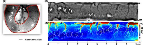
Using an experimental NEC model, we acquired in vivo images of the gut microvasculature and blood flow over a 5000 × 7000 μm2 field of view at 5 μm resolution via the following two endogenous contrast mechanisms: intrinsic optical signals and laser speckles. Next, we transformed intestinal images into rectilinear “flat maps,” and delineated 1A/V gut microvessels and their perfusion territories as “intestinal vascular units” (IVUs). Employing IVUs, we quantified and visualized NEC-induced changes to the gut vascular phenotype.
Results
In vivo imaging required 60–100 s per animal. Relative to the healthy gut, NEC intestines showed a significant overall decrease (i.e. 64–72%) in perfusion, accompanied by vasoconstriction (i.e. 9–12%) and a reduction in perfusion entropy (19%)within sections of the vascular bed.
Conclusions
Multicontrast imaging coupled with IVU-based in vivo vascular phenotyping is a powerful new tool for elucidating NEC pathogenesis.
期刊介绍:
The journal features original contributions that are the result of investigations contributing significant new information relating to the vascular and lymphatic microcirculation addressed at the intact animal, organ, cellular, or molecular level. Papers describe applications of the methods of physiology, biophysics, bioengineering, genetics, cell biology, biochemistry, and molecular biology to problems in microcirculation.
Microcirculation also publishes state-of-the-art reviews that address frontier areas or new advances in technology in the fields of microcirculatory disease and function. Specific areas of interest include: Angiogenesis, growth and remodeling; Transport and exchange of gasses and solutes; Rheology and biorheology; Endothelial cell biology and metabolism; Interactions between endothelium, smooth muscle, parenchymal cells, leukocytes and platelets; Regulation of vasomotor tone; and Microvascular structures, imaging and morphometry. Papers also describe innovations in experimental techniques and instrumentation for studying all aspects of microcirculatory structure and function.
 求助内容:
求助内容: 应助结果提醒方式:


