Deniz Kavzak Ufuktepe, Feng Yang, Yasmin M Kassim, Hang Yu, Richard J Maude, Kannappan Palaniappan, Stefan Jaeger
{"title":"基于深度学习的细胞检测与提取,用于疟疾诊断的薄血涂片。","authors":"Deniz Kavzak Ufuktepe, Feng Yang, Yasmin M Kassim, Hang Yu, Richard J Maude, Kannappan Palaniappan, Stefan Jaeger","doi":"10.1109/AIPR52630.2021.9762109","DOIUrl":null,"url":null,"abstract":"<p><p>Malaria is a major health threat caused by Plasmodium parasites that infect the red blood cells. Two predominant types of Plasmodium parasites are <i>Plasmodium vivax</i> (<i>P</i>. <i>vivax</i>) and <i>Plasmodium falciparum</i> (<i>P</i>. <i>falciparum</i>). Diagnosis of malaria typically involves visual microscopy examination of blood smears for malaria parasites. This is a tedious, error-prone visual inspection task requiring microscopy expertise which is often lacking in resource-poor settings. To address these problems, attempts have been made in recent years to automate malaria diagnosis using machine learning approaches. Several challenges need to be met for a machine learning approach to be successful in malaria diagnosis. Microscopy images acquired at different sites often vary in color, contrast, and consistency caused by different smear preparation and staining methods. Moreover, touching and overlapping cells complicate the red blood cell detection process, which can lead to inaccurate blood cell counts and thus incorrect parasitemia calculations. In this work, we propose a red blood cell detection and extraction framework to enable processing and analysis of single cells for follow-up processes like counting infected cells or identifying parasite species in thin blood smears. This framework consists of two modules: a cell detection module and a cell extraction module. The cell detection module trains a modified Channel-wise Feature Pyramid Network for Medicine (CFPNet-M) deep learning network that takes the green channel of the image and the color-deconvolution processed image as inputs, and learns a truncated distance transform image of cell annotations. CFPNet-M is chosen due to its low resource requirements, while the distance transform allows achieving more accurate cell counts for dense cells. Once the cells are detected by the network, the cell extraction module is used to extract single cells from the original image and count the number of cells. Our preliminary results based on 193 patients (including 148 <i>P</i>. <i>Falciparum</i> infected patients, and 45 uninfected patients) show that our framework achieves cell count accuracy of 92.2%.</p>","PeriodicalId":73278,"journal":{"name":"IEEE Applied Imagery Pattern Recognition Workshop : [proceedings]. IEEE Applied Imagery Pattern Recognition Workshop","volume":"2021 ","pages":"9762109"},"PeriodicalIF":0.0000,"publicationDate":"2021-04-26","publicationTypes":"Journal Article","fieldsOfStudy":null,"isOpenAccess":false,"openAccessPdf":"https://www.ncbi.nlm.nih.gov/pmc/articles/PMC7613898/pdf/","citationCount":"0","resultStr":"{\"title\":\"Deep Learning-Based Cell Detection and Extraction in Thin Blood Smears for Malaria Diagnosis.\",\"authors\":\"Deniz Kavzak Ufuktepe, Feng Yang, Yasmin M Kassim, Hang Yu, Richard J Maude, Kannappan Palaniappan, Stefan Jaeger\",\"doi\":\"10.1109/AIPR52630.2021.9762109\",\"DOIUrl\":null,\"url\":null,\"abstract\":\"<p><p>Malaria is a major health threat caused by Plasmodium parasites that infect the red blood cells. Two predominant types of Plasmodium parasites are <i>Plasmodium vivax</i> (<i>P</i>. <i>vivax</i>) and <i>Plasmodium falciparum</i> (<i>P</i>. <i>falciparum</i>). Diagnosis of malaria typically involves visual microscopy examination of blood smears for malaria parasites. This is a tedious, error-prone visual inspection task requiring microscopy expertise which is often lacking in resource-poor settings. To address these problems, attempts have been made in recent years to automate malaria diagnosis using machine learning approaches. Several challenges need to be met for a machine learning approach to be successful in malaria diagnosis. Microscopy images acquired at different sites often vary in color, contrast, and consistency caused by different smear preparation and staining methods. Moreover, touching and overlapping cells complicate the red blood cell detection process, which can lead to inaccurate blood cell counts and thus incorrect parasitemia calculations. In this work, we propose a red blood cell detection and extraction framework to enable processing and analysis of single cells for follow-up processes like counting infected cells or identifying parasite species in thin blood smears. This framework consists of two modules: a cell detection module and a cell extraction module. The cell detection module trains a modified Channel-wise Feature Pyramid Network for Medicine (CFPNet-M) deep learning network that takes the green channel of the image and the color-deconvolution processed image as inputs, and learns a truncated distance transform image of cell annotations. CFPNet-M is chosen due to its low resource requirements, while the distance transform allows achieving more accurate cell counts for dense cells. Once the cells are detected by the network, the cell extraction module is used to extract single cells from the original image and count the number of cells. Our preliminary results based on 193 patients (including 148 <i>P</i>. <i>Falciparum</i> infected patients, and 45 uninfected patients) show that our framework achieves cell count accuracy of 92.2%.</p>\",\"PeriodicalId\":73278,\"journal\":{\"name\":\"IEEE Applied Imagery Pattern Recognition Workshop : [proceedings]. IEEE Applied Imagery Pattern Recognition Workshop\",\"volume\":\"2021 \",\"pages\":\"9762109\"},\"PeriodicalIF\":0.0000,\"publicationDate\":\"2021-04-26\",\"publicationTypes\":\"Journal Article\",\"fieldsOfStudy\":null,\"isOpenAccess\":false,\"openAccessPdf\":\"https://www.ncbi.nlm.nih.gov/pmc/articles/PMC7613898/pdf/\",\"citationCount\":\"0\",\"resultStr\":null,\"platform\":\"Semanticscholar\",\"paperid\":null,\"PeriodicalName\":\"IEEE Applied Imagery Pattern Recognition Workshop : [proceedings]. IEEE Applied Imagery Pattern Recognition Workshop\",\"FirstCategoryId\":\"1085\",\"ListUrlMain\":\"https://doi.org/10.1109/AIPR52630.2021.9762109\",\"RegionNum\":0,\"RegionCategory\":null,\"ArticlePicture\":[],\"TitleCN\":null,\"AbstractTextCN\":null,\"PMCID\":null,\"EPubDate\":\"\",\"PubModel\":\"\",\"JCR\":\"\",\"JCRName\":\"\",\"Score\":null,\"Total\":0}","platform":"Semanticscholar","paperid":null,"PeriodicalName":"IEEE Applied Imagery Pattern Recognition Workshop : [proceedings]. IEEE Applied Imagery Pattern Recognition Workshop","FirstCategoryId":"1085","ListUrlMain":"https://doi.org/10.1109/AIPR52630.2021.9762109","RegionNum":0,"RegionCategory":null,"ArticlePicture":[],"TitleCN":null,"AbstractTextCN":null,"PMCID":null,"EPubDate":"","PubModel":"","JCR":"","JCRName":"","Score":null,"Total":0}
Deep Learning-Based Cell Detection and Extraction in Thin Blood Smears for Malaria Diagnosis.
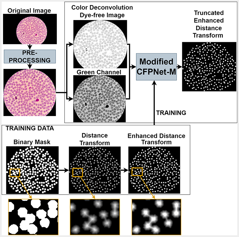
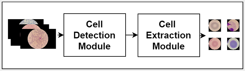
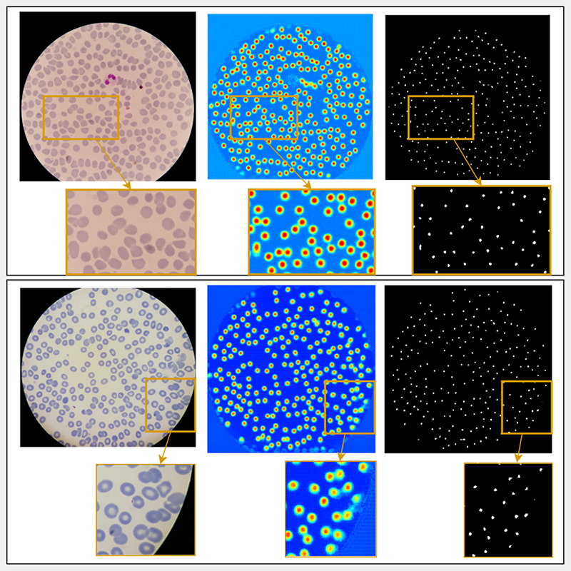
Malaria is a major health threat caused by Plasmodium parasites that infect the red blood cells. Two predominant types of Plasmodium parasites are Plasmodium vivax (P. vivax) and Plasmodium falciparum (P. falciparum). Diagnosis of malaria typically involves visual microscopy examination of blood smears for malaria parasites. This is a tedious, error-prone visual inspection task requiring microscopy expertise which is often lacking in resource-poor settings. To address these problems, attempts have been made in recent years to automate malaria diagnosis using machine learning approaches. Several challenges need to be met for a machine learning approach to be successful in malaria diagnosis. Microscopy images acquired at different sites often vary in color, contrast, and consistency caused by different smear preparation and staining methods. Moreover, touching and overlapping cells complicate the red blood cell detection process, which can lead to inaccurate blood cell counts and thus incorrect parasitemia calculations. In this work, we propose a red blood cell detection and extraction framework to enable processing and analysis of single cells for follow-up processes like counting infected cells or identifying parasite species in thin blood smears. This framework consists of two modules: a cell detection module and a cell extraction module. The cell detection module trains a modified Channel-wise Feature Pyramid Network for Medicine (CFPNet-M) deep learning network that takes the green channel of the image and the color-deconvolution processed image as inputs, and learns a truncated distance transform image of cell annotations. CFPNet-M is chosen due to its low resource requirements, while the distance transform allows achieving more accurate cell counts for dense cells. Once the cells are detected by the network, the cell extraction module is used to extract single cells from the original image and count the number of cells. Our preliminary results based on 193 patients (including 148 P. Falciparum infected patients, and 45 uninfected patients) show that our framework achieves cell count accuracy of 92.2%.
![IEEE Applied Imagery Pattern Recognition Workshop : [proceedings]. IEEE Applied Imagery Pattern Recognition Workshop](/Content/css/sci/img/zetp.jpg)
 求助内容:
求助内容: 应助结果提醒方式:


