{"title":"木材防腐剂铬化砷酸铜暴露4周后大鼠肝脏抗氧化和DNA甲基化基因表达分析。","authors":"Naofumi Takahashi, Satoru Yamaguchi, Ryouichi Ohtsuka, Makio Takeda, Toshinori Yoshida, Tadashi Kosaka, Takanori Harada","doi":"10.1293/tox.2022-0093","DOIUrl":null,"url":null,"abstract":"<p><p>Our previous 4-week repeated dose toxicity study showed that wood preservative chromated copper arsenate (CCA) induced hepatocellular hypertrophy accompanied by biochemical hepatic dysfunction and an increase in oxidative stress marker, 8-hydroxydeoxyguanosine, in female rats. To further explore the molecular mechanisms of CCA hepatotoxicity, we analyzed 10%-buffered formalin-fixed liver samples from female rats for cell proliferation, apoptosis, and protein glutathionylation and conducted microarray analysis on frozen liver samples from female rats treated with 0 or 80 mg/kg/day of CCA. Chemical analysis revealed that dimethylated arsenical was the major metabolite in liver tissues of male and female rats. CCA increase labeling indices of proliferating cell nuclear antigen and decrease terminal deoxynucleotidyl transferase-mediated dUTP nick-end labeling accompanied with increased expression of protein glutathionylation, indicating a decrease in glutathione (GSH) in hepatocytes of female rats. Microarray analysis revealed that CCA altered gene expression of antioxidants, glutathione-S-transferase (GST), heat shock proteins and ubiquitin-proteasome pathway, cell proliferation, apoptosis, DNA methylation, cytochrome P450, and glucose and lipid metabolism in female rats. Increased expression of GSTs, including <i>Gsta2</i>, <i>Gsta3</i>, <i>Mgst1</i>, and <i>Cdkn1b</i> (<i>p27</i>), and decreased expression of the antioxidant <i>Mt1</i>, and DNA methylation <i>Dnmt1</i>, <i>Dnmt3a</i>, and <i>Ctcf</i> were confirmed in the liver of female rats in a dose-dependent manner. Methylation status of the promoter region of the <i>Mt1</i> was not evidently changed between control and treatment groups. The results suggested that CCA decreased GSH and altered the expression of several genes, including antioxidants, GST, and DNA methylation, followed by impaired cell proliferation in the liver of female rats.</p>","PeriodicalId":17437,"journal":{"name":"Journal of Toxicologic Pathology","volume":"36 1","pages":"31-43"},"PeriodicalIF":0.9000,"publicationDate":"2023-01-01","publicationTypes":"Journal Article","fieldsOfStudy":null,"isOpenAccess":false,"openAccessPdf":"https://ftp.ncbi.nlm.nih.gov/pub/pmc/oa_pdf/e4/80/tox-36-031.PMC9837468.pdf","citationCount":"0","resultStr":"{\"title\":\"Gene expression analysis of antioxidant and DNA methylation on the rat liver after 4-week wood preservative chromated copper arsenate exposure.\",\"authors\":\"Naofumi Takahashi, Satoru Yamaguchi, Ryouichi Ohtsuka, Makio Takeda, Toshinori Yoshida, Tadashi Kosaka, Takanori Harada\",\"doi\":\"10.1293/tox.2022-0093\",\"DOIUrl\":null,\"url\":null,\"abstract\":\"<p><p>Our previous 4-week repeated dose toxicity study showed that wood preservative chromated copper arsenate (CCA) induced hepatocellular hypertrophy accompanied by biochemical hepatic dysfunction and an increase in oxidative stress marker, 8-hydroxydeoxyguanosine, in female rats. To further explore the molecular mechanisms of CCA hepatotoxicity, we analyzed 10%-buffered formalin-fixed liver samples from female rats for cell proliferation, apoptosis, and protein glutathionylation and conducted microarray analysis on frozen liver samples from female rats treated with 0 or 80 mg/kg/day of CCA. Chemical analysis revealed that dimethylated arsenical was the major metabolite in liver tissues of male and female rats. CCA increase labeling indices of proliferating cell nuclear antigen and decrease terminal deoxynucleotidyl transferase-mediated dUTP nick-end labeling accompanied with increased expression of protein glutathionylation, indicating a decrease in glutathione (GSH) in hepatocytes of female rats. Microarray analysis revealed that CCA altered gene expression of antioxidants, glutathione-S-transferase (GST), heat shock proteins and ubiquitin-proteasome pathway, cell proliferation, apoptosis, DNA methylation, cytochrome P450, and glucose and lipid metabolism in female rats. Increased expression of GSTs, including <i>Gsta2</i>, <i>Gsta3</i>, <i>Mgst1</i>, and <i>Cdkn1b</i> (<i>p27</i>), and decreased expression of the antioxidant <i>Mt1</i>, and DNA methylation <i>Dnmt1</i>, <i>Dnmt3a</i>, and <i>Ctcf</i> were confirmed in the liver of female rats in a dose-dependent manner. Methylation status of the promoter region of the <i>Mt1</i> was not evidently changed between control and treatment groups. The results suggested that CCA decreased GSH and altered the expression of several genes, including antioxidants, GST, and DNA methylation, followed by impaired cell proliferation in the liver of female rats.</p>\",\"PeriodicalId\":17437,\"journal\":{\"name\":\"Journal of Toxicologic Pathology\",\"volume\":\"36 1\",\"pages\":\"31-43\"},\"PeriodicalIF\":0.9000,\"publicationDate\":\"2023-01-01\",\"publicationTypes\":\"Journal Article\",\"fieldsOfStudy\":null,\"isOpenAccess\":false,\"openAccessPdf\":\"https://ftp.ncbi.nlm.nih.gov/pub/pmc/oa_pdf/e4/80/tox-36-031.PMC9837468.pdf\",\"citationCount\":\"0\",\"resultStr\":null,\"platform\":\"Semanticscholar\",\"paperid\":null,\"PeriodicalName\":\"Journal of Toxicologic Pathology\",\"FirstCategoryId\":\"3\",\"ListUrlMain\":\"https://doi.org/10.1293/tox.2022-0093\",\"RegionNum\":4,\"RegionCategory\":\"医学\",\"ArticlePicture\":[],\"TitleCN\":null,\"AbstractTextCN\":null,\"PMCID\":null,\"EPubDate\":\"\",\"PubModel\":\"\",\"JCR\":\"Q4\",\"JCRName\":\"PATHOLOGY\",\"Score\":null,\"Total\":0}","platform":"Semanticscholar","paperid":null,"PeriodicalName":"Journal of Toxicologic Pathology","FirstCategoryId":"3","ListUrlMain":"https://doi.org/10.1293/tox.2022-0093","RegionNum":4,"RegionCategory":"医学","ArticlePicture":[],"TitleCN":null,"AbstractTextCN":null,"PMCID":null,"EPubDate":"","PubModel":"","JCR":"Q4","JCRName":"PATHOLOGY","Score":null,"Total":0}
Gene expression analysis of antioxidant and DNA methylation on the rat liver after 4-week wood preservative chromated copper arsenate exposure.
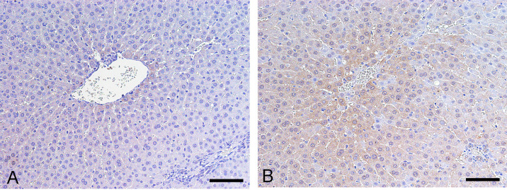
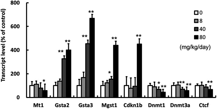
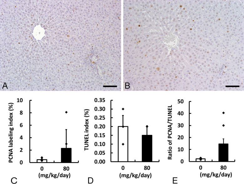
Our previous 4-week repeated dose toxicity study showed that wood preservative chromated copper arsenate (CCA) induced hepatocellular hypertrophy accompanied by biochemical hepatic dysfunction and an increase in oxidative stress marker, 8-hydroxydeoxyguanosine, in female rats. To further explore the molecular mechanisms of CCA hepatotoxicity, we analyzed 10%-buffered formalin-fixed liver samples from female rats for cell proliferation, apoptosis, and protein glutathionylation and conducted microarray analysis on frozen liver samples from female rats treated with 0 or 80 mg/kg/day of CCA. Chemical analysis revealed that dimethylated arsenical was the major metabolite in liver tissues of male and female rats. CCA increase labeling indices of proliferating cell nuclear antigen and decrease terminal deoxynucleotidyl transferase-mediated dUTP nick-end labeling accompanied with increased expression of protein glutathionylation, indicating a decrease in glutathione (GSH) in hepatocytes of female rats. Microarray analysis revealed that CCA altered gene expression of antioxidants, glutathione-S-transferase (GST), heat shock proteins and ubiquitin-proteasome pathway, cell proliferation, apoptosis, DNA methylation, cytochrome P450, and glucose and lipid metabolism in female rats. Increased expression of GSTs, including Gsta2, Gsta3, Mgst1, and Cdkn1b (p27), and decreased expression of the antioxidant Mt1, and DNA methylation Dnmt1, Dnmt3a, and Ctcf were confirmed in the liver of female rats in a dose-dependent manner. Methylation status of the promoter region of the Mt1 was not evidently changed between control and treatment groups. The results suggested that CCA decreased GSH and altered the expression of several genes, including antioxidants, GST, and DNA methylation, followed by impaired cell proliferation in the liver of female rats.
期刊介绍:
JTP is a scientific journal that publishes original studies in the field of toxicological pathology and in a wide variety of other related fields. The main scope of the journal is listed below.
Administrative Opinions of Policymakers and Regulatory Agencies
Adverse Events
Carcinogenesis
Data of A Predominantly Negative Nature
Drug-Induced Hematologic Toxicity
Embryological Pathology
High Throughput Pathology
Historical Data of Experimental Animals
Immunohistochemical Analysis
Molecular Pathology
Nomenclature of Lesions
Non-mammal Toxicity Study
Result or Lesion Induced by Chemicals of Which Names Hidden on Account of the Authors
Technology and Methodology Related to Toxicological Pathology
Tumor Pathology; Neoplasia and Hyperplasia
Ultrastructural Analysis
Use of Animal Models.
 求助内容:
求助内容: 应助结果提醒方式:


