Alexander Kapustin, Sofia Serena Tsakali, Meredith Whitehead, George Chennell, Meng-Ying Wu, Chris Molenaar, Anton Kutikhin, Yimeng Chen, Sadia Ahmad, Leo Bogdanov, Maxim Sinitsky, Kseniya Rubina, Aled Clayton, Frederik J Verweij, Dirk Michiel Pegtel, Simona Zingaro, Arseniy Lobov, Bozhana Zainullina, Dylan Owen, Maddy Parsons, Richard E Cheney, Derek Warren, Martin James Humphries, Thomas Iskratsch, Mark Holt, Catherine M Shanahan
{"title":"细胞外小泡通过呈递胶原VI刺激平滑肌细胞迁移。","authors":"Alexander Kapustin, Sofia Serena Tsakali, Meredith Whitehead, George Chennell, Meng-Ying Wu, Chris Molenaar, Anton Kutikhin, Yimeng Chen, Sadia Ahmad, Leo Bogdanov, Maxim Sinitsky, Kseniya Rubina, Aled Clayton, Frederik J Verweij, Dirk Michiel Pegtel, Simona Zingaro, Arseniy Lobov, Bozhana Zainullina, Dylan Owen, Maddy Parsons, Richard E Cheney, Derek Warren, Martin James Humphries, Thomas Iskratsch, Mark Holt, Catherine M Shanahan","doi":"10.1101/2023.08.17.551257","DOIUrl":null,"url":null,"abstract":"<p><p>The extracellular matrix (ECM) supports blood vessel architecture and functionality and undergoes active remodelling during vascular repair and atherogenesis. Vascular smooth muscle cells (VSMCs) are essential for vessel repair and, via their secretome, can invade from the vessel media into the intima to mediate ECM remodelling. Accumulation of fibronectin (FN) is a hallmark of early vascular repair and atherosclerosis. Here we show that FN stimulates VSMCs to secrete small extracellular vesicles (sEVs) by activating the β1 integrin/FAK/Src pathway as well as Arp2/3-dependent branching of the actin cytoskeleton. We found that sEVs are trapped by the ECM in vitro and colocalise with FN in symptomatic atherosclerotic plaques in vivo. Functionally, ECM-trapped sEVs induced the formation of focal adhesions (FA) with enhanced pulling forces at the cellular periphery preventing cellular spreading and adhesion. Proteomic and GO pathway analysis revealed that VSMC-derived sEVs display a cell adhesion signature and are specifically enriched with collagen VI on the sEV surface. In vitro assays identified collagen VI as playing a key role in cell adhesion and invasion directionality. Taken together our data suggests that the accumulation of FN is a key early event in vessel repair acting to promote secretion of collage VI enriched sEVs by VSMCs. These sEVs stimulate directional invasion, most likely by triggering peripheral focal adhesion formation and actomyosin contraction to exert sufficient traction force to enable VSMC movement within the complex vascular ECM network.</p><p><strong>Figure abstract: </strong>Vascular smooth muscle cells sense fibronectin via β1 integrin and secrete small extracellular vesicles loaded with collagen VI. These extracellular vesicles are entrapped in the extracellular matrix and induce formation of peripheral focal adhesions presenting adhesion complex ECM proteins including collagen VI, LGALS3BP, EDIL3 and TGFBI. Focal adhesions anchor the extracellular matrix to actin fibrils in the cell. Contraction of the actin fibrils generates the mechanical force for directional cell invasion through the matrix. This figure was created with BioRender ( https://biorender.com/ ).</p>","PeriodicalId":72407,"journal":{"name":"bioRxiv : the preprint server for biology","volume":" ","pages":""},"PeriodicalIF":0.0000,"publicationDate":"2025-07-10","publicationTypes":"Journal Article","fieldsOfStudy":null,"isOpenAccess":false,"openAccessPdf":"https://www.ncbi.nlm.nih.gov/pmc/articles/PMC10462164/pdf/","citationCount":"0","resultStr":"{\"title\":\"Matrix-associated extracellular vesicles modulate smooth muscle cell adhesion and directionality by presenting collagen VI.\",\"authors\":\"Alexander Kapustin, Sofia Serena Tsakali, Meredith Whitehead, George Chennell, Meng-Ying Wu, Chris Molenaar, Anton Kutikhin, Yimeng Chen, Sadia Ahmad, Leo Bogdanov, Maxim Sinitsky, Kseniya Rubina, Aled Clayton, Frederik J Verweij, Dirk Michiel Pegtel, Simona Zingaro, Arseniy Lobov, Bozhana Zainullina, Dylan Owen, Maddy Parsons, Richard E Cheney, Derek Warren, Martin James Humphries, Thomas Iskratsch, Mark Holt, Catherine M Shanahan\",\"doi\":\"10.1101/2023.08.17.551257\",\"DOIUrl\":null,\"url\":null,\"abstract\":\"<p><p>The extracellular matrix (ECM) supports blood vessel architecture and functionality and undergoes active remodelling during vascular repair and atherogenesis. Vascular smooth muscle cells (VSMCs) are essential for vessel repair and, via their secretome, can invade from the vessel media into the intima to mediate ECM remodelling. Accumulation of fibronectin (FN) is a hallmark of early vascular repair and atherosclerosis. Here we show that FN stimulates VSMCs to secrete small extracellular vesicles (sEVs) by activating the β1 integrin/FAK/Src pathway as well as Arp2/3-dependent branching of the actin cytoskeleton. We found that sEVs are trapped by the ECM in vitro and colocalise with FN in symptomatic atherosclerotic plaques in vivo. Functionally, ECM-trapped sEVs induced the formation of focal adhesions (FA) with enhanced pulling forces at the cellular periphery preventing cellular spreading and adhesion. Proteomic and GO pathway analysis revealed that VSMC-derived sEVs display a cell adhesion signature and are specifically enriched with collagen VI on the sEV surface. In vitro assays identified collagen VI as playing a key role in cell adhesion and invasion directionality. Taken together our data suggests that the accumulation of FN is a key early event in vessel repair acting to promote secretion of collage VI enriched sEVs by VSMCs. These sEVs stimulate directional invasion, most likely by triggering peripheral focal adhesion formation and actomyosin contraction to exert sufficient traction force to enable VSMC movement within the complex vascular ECM network.</p><p><strong>Figure abstract: </strong>Vascular smooth muscle cells sense fibronectin via β1 integrin and secrete small extracellular vesicles loaded with collagen VI. These extracellular vesicles are entrapped in the extracellular matrix and induce formation of peripheral focal adhesions presenting adhesion complex ECM proteins including collagen VI, LGALS3BP, EDIL3 and TGFBI. Focal adhesions anchor the extracellular matrix to actin fibrils in the cell. Contraction of the actin fibrils generates the mechanical force for directional cell invasion through the matrix. This figure was created with BioRender ( https://biorender.com/ ).</p>\",\"PeriodicalId\":72407,\"journal\":{\"name\":\"bioRxiv : the preprint server for biology\",\"volume\":\" \",\"pages\":\"\"},\"PeriodicalIF\":0.0000,\"publicationDate\":\"2025-07-10\",\"publicationTypes\":\"Journal Article\",\"fieldsOfStudy\":null,\"isOpenAccess\":false,\"openAccessPdf\":\"https://www.ncbi.nlm.nih.gov/pmc/articles/PMC10462164/pdf/\",\"citationCount\":\"0\",\"resultStr\":null,\"platform\":\"Semanticscholar\",\"paperid\":null,\"PeriodicalName\":\"bioRxiv : the preprint server for biology\",\"FirstCategoryId\":\"1085\",\"ListUrlMain\":\"https://doi.org/10.1101/2023.08.17.551257\",\"RegionNum\":0,\"RegionCategory\":null,\"ArticlePicture\":[],\"TitleCN\":null,\"AbstractTextCN\":null,\"PMCID\":null,\"EPubDate\":\"\",\"PubModel\":\"\",\"JCR\":\"\",\"JCRName\":\"\",\"Score\":null,\"Total\":0}","platform":"Semanticscholar","paperid":null,"PeriodicalName":"bioRxiv : the preprint server for biology","FirstCategoryId":"1085","ListUrlMain":"https://doi.org/10.1101/2023.08.17.551257","RegionNum":0,"RegionCategory":null,"ArticlePicture":[],"TitleCN":null,"AbstractTextCN":null,"PMCID":null,"EPubDate":"","PubModel":"","JCR":"","JCRName":"","Score":null,"Total":0}
Matrix-associated extracellular vesicles modulate smooth muscle cell adhesion and directionality by presenting collagen VI.
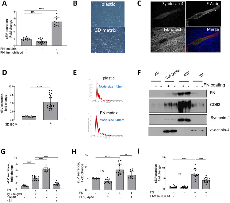
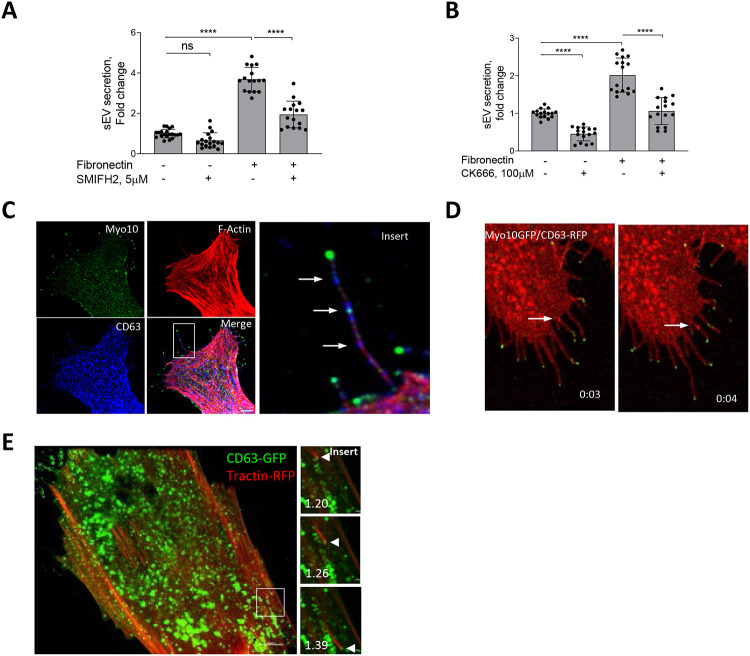
The extracellular matrix (ECM) supports blood vessel architecture and functionality and undergoes active remodelling during vascular repair and atherogenesis. Vascular smooth muscle cells (VSMCs) are essential for vessel repair and, via their secretome, can invade from the vessel media into the intima to mediate ECM remodelling. Accumulation of fibronectin (FN) is a hallmark of early vascular repair and atherosclerosis. Here we show that FN stimulates VSMCs to secrete small extracellular vesicles (sEVs) by activating the β1 integrin/FAK/Src pathway as well as Arp2/3-dependent branching of the actin cytoskeleton. We found that sEVs are trapped by the ECM in vitro and colocalise with FN in symptomatic atherosclerotic plaques in vivo. Functionally, ECM-trapped sEVs induced the formation of focal adhesions (FA) with enhanced pulling forces at the cellular periphery preventing cellular spreading and adhesion. Proteomic and GO pathway analysis revealed that VSMC-derived sEVs display a cell adhesion signature and are specifically enriched with collagen VI on the sEV surface. In vitro assays identified collagen VI as playing a key role in cell adhesion and invasion directionality. Taken together our data suggests that the accumulation of FN is a key early event in vessel repair acting to promote secretion of collage VI enriched sEVs by VSMCs. These sEVs stimulate directional invasion, most likely by triggering peripheral focal adhesion formation and actomyosin contraction to exert sufficient traction force to enable VSMC movement within the complex vascular ECM network.
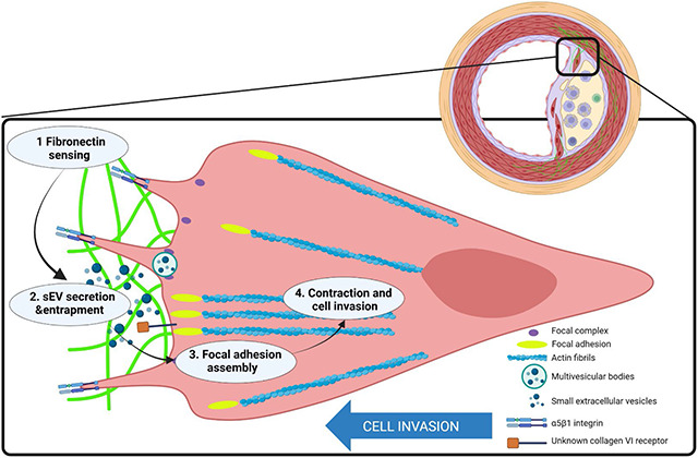
Figure abstract: Vascular smooth muscle cells sense fibronectin via β1 integrin and secrete small extracellular vesicles loaded with collagen VI. These extracellular vesicles are entrapped in the extracellular matrix and induce formation of peripheral focal adhesions presenting adhesion complex ECM proteins including collagen VI, LGALS3BP, EDIL3 and TGFBI. Focal adhesions anchor the extracellular matrix to actin fibrils in the cell. Contraction of the actin fibrils generates the mechanical force for directional cell invasion through the matrix. This figure was created with BioRender ( https://biorender.com/ ).

 求助内容:
求助内容: 应助结果提醒方式:


