{"title":"使用非增强磁共振成像识别右肾上腺静脉。","authors":"Koji Yamashita, Noriaki Wada, Seitaro Shin, Tetsuhiro Watanabe, Kiyomi Furuya, Shino Harada, Tomoyuki Noguchi","doi":"10.5114/pjr.2022.121236","DOIUrl":null,"url":null,"abstract":"<p><strong>Purpose: </strong>Unenhanced magnetic resonance imaging (MRI) is known to be useful in characterizing adrenal adenomas through the implementation of in-phase (IPI) and opposed-phase imaging (OPI) based on chemical shift artifacts. However, whether unenhanced MRI can contribute to the identification of right adrenal vein (RAV) remains unclear. The aim of this study was to evaluate the feasibility of unenhanced MRI for the identification of RAV.</p><p><strong>Material and methods: </strong>This retrospective study reviewed 30 patients (16 men; median age 60 years; range 34-76 years) who underwent MRI and subsequent adrenal venous sampling (AVS). Chemical shift MRI was acquired using echo times of 2.3 ms (OPI) and 4.6 ms (IPI) with a slice thickness of 3 mm and a gap of 1 mm. T2-weighted imaging (T2WI) was also performed. Identification of RAVs was performed by 2 independent radiologists. Inter-observer agreement on a 3-point rating scale was evaluated using κ statistics. The identification rate of RAVs was compared between OPI, IPI, and T2WI using McNemar's test.</p><p><strong>Results: </strong>Good inter-observer agreement was found for the OPI (κ = 0.744), whereas fair agreement was obtained for both other sequences (IPI: κ = 0.375; T2WI: 0.348). For both raters, the identification rate of RAVs was higher with OPI (36/60; 60.0%) than with other sequences (IPI: 16/60, 26.7%; T2WI: 9/60, 15.0%; <i>p</i> < 0.05, each).</p><p><strong>Conclusions: </strong>OPI may play a screening role in the identification of RAVs preceding AVS, which could reduce the required radiation exposure and doses of contrast agent.</p>","PeriodicalId":47128,"journal":{"name":"Polish Journal of Radiology","volume":"87 ","pages":"e592-e596"},"PeriodicalIF":1.6000,"publicationDate":"2022-01-01","publicationTypes":"Journal Article","fieldsOfStudy":null,"isOpenAccess":false,"openAccessPdf":"https://ftp.ncbi.nlm.nih.gov/pub/pmc/oa_pdf/ff/7e/PJR-87-48193.PMC9749782.pdf","citationCount":"0","resultStr":"{\"title\":\"Right adrenal vein identification using unenhanced magnetic resonance imaging.\",\"authors\":\"Koji Yamashita, Noriaki Wada, Seitaro Shin, Tetsuhiro Watanabe, Kiyomi Furuya, Shino Harada, Tomoyuki Noguchi\",\"doi\":\"10.5114/pjr.2022.121236\",\"DOIUrl\":null,\"url\":null,\"abstract\":\"<p><strong>Purpose: </strong>Unenhanced magnetic resonance imaging (MRI) is known to be useful in characterizing adrenal adenomas through the implementation of in-phase (IPI) and opposed-phase imaging (OPI) based on chemical shift artifacts. However, whether unenhanced MRI can contribute to the identification of right adrenal vein (RAV) remains unclear. The aim of this study was to evaluate the feasibility of unenhanced MRI for the identification of RAV.</p><p><strong>Material and methods: </strong>This retrospective study reviewed 30 patients (16 men; median age 60 years; range 34-76 years) who underwent MRI and subsequent adrenal venous sampling (AVS). Chemical shift MRI was acquired using echo times of 2.3 ms (OPI) and 4.6 ms (IPI) with a slice thickness of 3 mm and a gap of 1 mm. T2-weighted imaging (T2WI) was also performed. Identification of RAVs was performed by 2 independent radiologists. Inter-observer agreement on a 3-point rating scale was evaluated using κ statistics. The identification rate of RAVs was compared between OPI, IPI, and T2WI using McNemar's test.</p><p><strong>Results: </strong>Good inter-observer agreement was found for the OPI (κ = 0.744), whereas fair agreement was obtained for both other sequences (IPI: κ = 0.375; T2WI: 0.348). For both raters, the identification rate of RAVs was higher with OPI (36/60; 60.0%) than with other sequences (IPI: 16/60, 26.7%; T2WI: 9/60, 15.0%; <i>p</i> < 0.05, each).</p><p><strong>Conclusions: </strong>OPI may play a screening role in the identification of RAVs preceding AVS, which could reduce the required radiation exposure and doses of contrast agent.</p>\",\"PeriodicalId\":47128,\"journal\":{\"name\":\"Polish Journal of Radiology\",\"volume\":\"87 \",\"pages\":\"e592-e596\"},\"PeriodicalIF\":1.6000,\"publicationDate\":\"2022-01-01\",\"publicationTypes\":\"Journal Article\",\"fieldsOfStudy\":null,\"isOpenAccess\":false,\"openAccessPdf\":\"https://ftp.ncbi.nlm.nih.gov/pub/pmc/oa_pdf/ff/7e/PJR-87-48193.PMC9749782.pdf\",\"citationCount\":\"0\",\"resultStr\":null,\"platform\":\"Semanticscholar\",\"paperid\":null,\"PeriodicalName\":\"Polish Journal of Radiology\",\"FirstCategoryId\":\"1085\",\"ListUrlMain\":\"https://doi.org/10.5114/pjr.2022.121236\",\"RegionNum\":0,\"RegionCategory\":null,\"ArticlePicture\":[],\"TitleCN\":null,\"AbstractTextCN\":null,\"PMCID\":null,\"EPubDate\":\"\",\"PubModel\":\"\",\"JCR\":\"Q4\",\"JCRName\":\"RADIOLOGY, NUCLEAR MEDICINE & MEDICAL IMAGING\",\"Score\":null,\"Total\":0}","platform":"Semanticscholar","paperid":null,"PeriodicalName":"Polish Journal of Radiology","FirstCategoryId":"1085","ListUrlMain":"https://doi.org/10.5114/pjr.2022.121236","RegionNum":0,"RegionCategory":null,"ArticlePicture":[],"TitleCN":null,"AbstractTextCN":null,"PMCID":null,"EPubDate":"","PubModel":"","JCR":"Q4","JCRName":"RADIOLOGY, NUCLEAR MEDICINE & MEDICAL IMAGING","Score":null,"Total":0}
引用次数: 0
摘要
目的:非增强磁共振成像(MRI)被认为是有用的,通过实施同相成像(IPI)和反相成像(OPI)基于化学位移伪影表征肾上腺腺瘤。然而,非增强MRI是否有助于识别右肾上腺静脉(RAV)仍不清楚。本研究的目的是评估非增强MRI识别RAV的可行性。材料和方法:本回顾性研究回顾了30例患者(16例男性;中位年龄60岁;范围34-76岁),接受MRI和随后的肾上腺静脉取样(AVS)。化学位移MRI的回波时间分别为2.3 ms (OPI)和4.6 ms (IPI),切片厚度为3 mm,间隙为1 mm。同时进行t2加权成像(T2WI)。RAVs的鉴定由2名独立放射科医生完成。使用κ统计量评估3点评定量表的观察者间一致性。采用McNemar试验比较OPI、IPI和T2WI对RAVs的检出率。结果:OPI的观察者间一致性较好(κ = 0.744),而其他两个序列的观察者间一致性较好(IPI: κ = 0.375;T2WI: 0.348)。对于两种评分者来说,OPI对RAVs的识别率更高(36/60;(IPI: 16/60, 26.7%;T2wi: 9/60, 15.0%;P < 0.05)。结论:OPI可能在AVS前的RAVs识别中起到筛选作用,可以减少所需的辐射暴露和造影剂剂量。
Right adrenal vein identification using unenhanced magnetic resonance imaging.
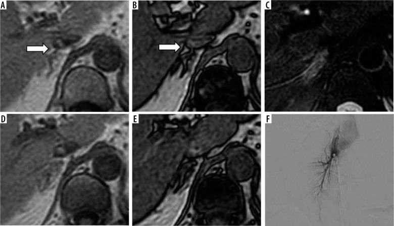
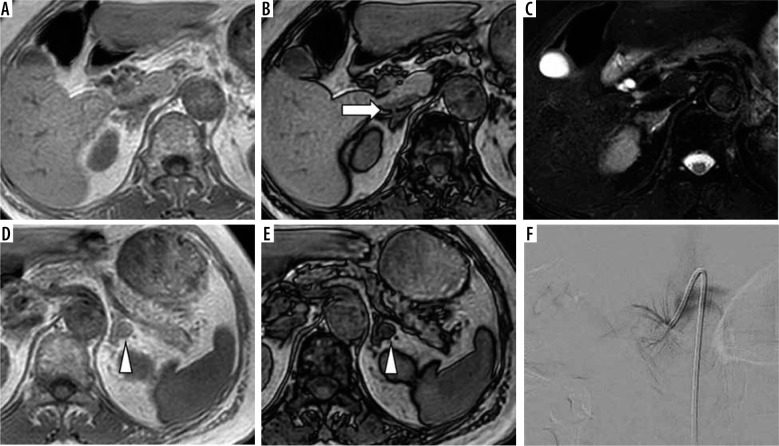
Purpose: Unenhanced magnetic resonance imaging (MRI) is known to be useful in characterizing adrenal adenomas through the implementation of in-phase (IPI) and opposed-phase imaging (OPI) based on chemical shift artifacts. However, whether unenhanced MRI can contribute to the identification of right adrenal vein (RAV) remains unclear. The aim of this study was to evaluate the feasibility of unenhanced MRI for the identification of RAV.
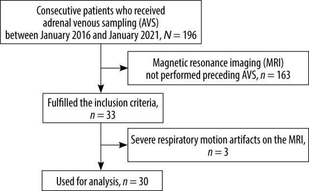
Material and methods: This retrospective study reviewed 30 patients (16 men; median age 60 years; range 34-76 years) who underwent MRI and subsequent adrenal venous sampling (AVS). Chemical shift MRI was acquired using echo times of 2.3 ms (OPI) and 4.6 ms (IPI) with a slice thickness of 3 mm and a gap of 1 mm. T2-weighted imaging (T2WI) was also performed. Identification of RAVs was performed by 2 independent radiologists. Inter-observer agreement on a 3-point rating scale was evaluated using κ statistics. The identification rate of RAVs was compared between OPI, IPI, and T2WI using McNemar's test.
Results: Good inter-observer agreement was found for the OPI (κ = 0.744), whereas fair agreement was obtained for both other sequences (IPI: κ = 0.375; T2WI: 0.348). For both raters, the identification rate of RAVs was higher with OPI (36/60; 60.0%) than with other sequences (IPI: 16/60, 26.7%; T2WI: 9/60, 15.0%; p < 0.05, each).
Conclusions: OPI may play a screening role in the identification of RAVs preceding AVS, which could reduce the required radiation exposure and doses of contrast agent.

 求助内容:
求助内容: 应助结果提醒方式:


