{"title":"全胃切除食管空肠造口术后食管空肠静脉曲张破裂1例经皮经肝闭塞双球囊封堵进、引流静脉成功。","authors":"Tsuyoshi Kawai, Shinsaku Yata, Kensuke Matsumoto, Kenichi Miyoshi, Naoya Noguchi, Shinya Fujii","doi":"10.22575/interventionalradiology.2022-0007","DOIUrl":null,"url":null,"abstract":"<p><p>We present the case of a man in his 60s with bleeding esophagojejunal varices occurring after gastrectomy for gastric carcinoma. Percutaneous transhepatic portography depicted the esophagojejunal varices originated from the jejunal vein and drained into the azygos vein. A 5-French occlusion balloon catheter was wedged into the jejunal vein and a 3-French occlusion balloon catheter into one drainage channel of the esophagojejunal varices via the azygos vein. Selective antegrade jejunal venography under dual-balloon occlusion revealed entire esophagojejunal varices with good stagnated and well-opacified contrast medium. Subsequently, 12 mL of 5% ethanolamine oleate-contrast medium mixture was slowly injected into the esophagojejunal varices. He was discharged without complications one week after the procedure, and abdominal computed tomography demonstrated the disappearance of the esophagojejunal varices six months after the procedure.</p>","PeriodicalId":73503,"journal":{"name":"Interventional radiology (Higashimatsuyama-shi (Japan)","volume":"7 3","pages":"114-118"},"PeriodicalIF":0.0000,"publicationDate":"2022-11-04","publicationTypes":"Journal Article","fieldsOfStudy":null,"isOpenAccess":false,"openAccessPdf":"https://ftp.ncbi.nlm.nih.gov/pub/pmc/oa_pdf/a8/3c/2432-0935-7-3-0114.PMC9719818.pdf","citationCount":"0","resultStr":"{\"title\":\"A Case of Esophagojejunal Variceal Rupture after Total Gastrectomy and Esophagojejunostomy Successfully Treated with Percutaneous Transhepatic Obliteration under Dual-balloon Occlusion of Feeding and Draining Veins.\",\"authors\":\"Tsuyoshi Kawai, Shinsaku Yata, Kensuke Matsumoto, Kenichi Miyoshi, Naoya Noguchi, Shinya Fujii\",\"doi\":\"10.22575/interventionalradiology.2022-0007\",\"DOIUrl\":null,\"url\":null,\"abstract\":\"<p><p>We present the case of a man in his 60s with bleeding esophagojejunal varices occurring after gastrectomy for gastric carcinoma. Percutaneous transhepatic portography depicted the esophagojejunal varices originated from the jejunal vein and drained into the azygos vein. A 5-French occlusion balloon catheter was wedged into the jejunal vein and a 3-French occlusion balloon catheter into one drainage channel of the esophagojejunal varices via the azygos vein. Selective antegrade jejunal venography under dual-balloon occlusion revealed entire esophagojejunal varices with good stagnated and well-opacified contrast medium. Subsequently, 12 mL of 5% ethanolamine oleate-contrast medium mixture was slowly injected into the esophagojejunal varices. He was discharged without complications one week after the procedure, and abdominal computed tomography demonstrated the disappearance of the esophagojejunal varices six months after the procedure.</p>\",\"PeriodicalId\":73503,\"journal\":{\"name\":\"Interventional radiology (Higashimatsuyama-shi (Japan)\",\"volume\":\"7 3\",\"pages\":\"114-118\"},\"PeriodicalIF\":0.0000,\"publicationDate\":\"2022-11-04\",\"publicationTypes\":\"Journal Article\",\"fieldsOfStudy\":null,\"isOpenAccess\":false,\"openAccessPdf\":\"https://ftp.ncbi.nlm.nih.gov/pub/pmc/oa_pdf/a8/3c/2432-0935-7-3-0114.PMC9719818.pdf\",\"citationCount\":\"0\",\"resultStr\":null,\"platform\":\"Semanticscholar\",\"paperid\":null,\"PeriodicalName\":\"Interventional radiology (Higashimatsuyama-shi (Japan)\",\"FirstCategoryId\":\"1085\",\"ListUrlMain\":\"https://doi.org/10.22575/interventionalradiology.2022-0007\",\"RegionNum\":0,\"RegionCategory\":null,\"ArticlePicture\":[],\"TitleCN\":null,\"AbstractTextCN\":null,\"PMCID\":null,\"EPubDate\":\"\",\"PubModel\":\"\",\"JCR\":\"\",\"JCRName\":\"\",\"Score\":null,\"Total\":0}","platform":"Semanticscholar","paperid":null,"PeriodicalName":"Interventional radiology (Higashimatsuyama-shi (Japan)","FirstCategoryId":"1085","ListUrlMain":"https://doi.org/10.22575/interventionalradiology.2022-0007","RegionNum":0,"RegionCategory":null,"ArticlePicture":[],"TitleCN":null,"AbstractTextCN":null,"PMCID":null,"EPubDate":"","PubModel":"","JCR":"","JCRName":"","Score":null,"Total":0}
A Case of Esophagojejunal Variceal Rupture after Total Gastrectomy and Esophagojejunostomy Successfully Treated with Percutaneous Transhepatic Obliteration under Dual-balloon Occlusion of Feeding and Draining Veins.
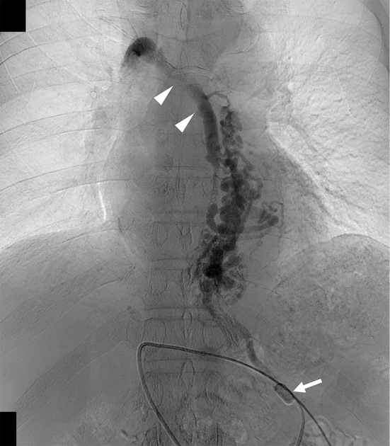
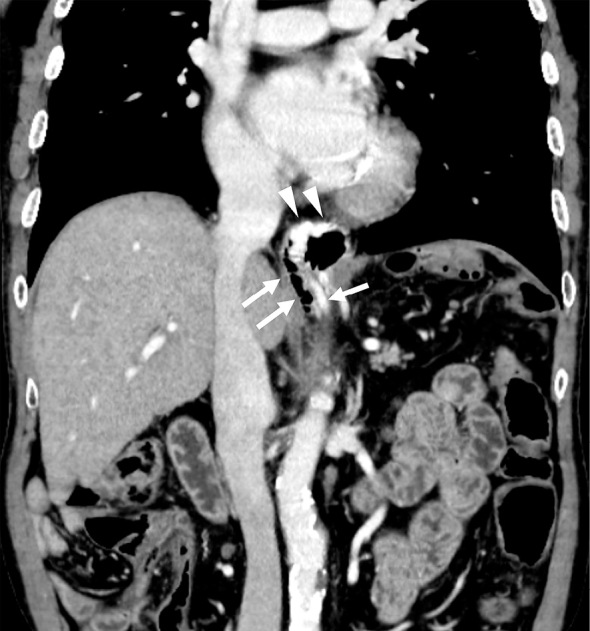
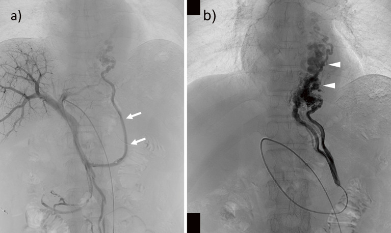
We present the case of a man in his 60s with bleeding esophagojejunal varices occurring after gastrectomy for gastric carcinoma. Percutaneous transhepatic portography depicted the esophagojejunal varices originated from the jejunal vein and drained into the azygos vein. A 5-French occlusion balloon catheter was wedged into the jejunal vein and a 3-French occlusion balloon catheter into one drainage channel of the esophagojejunal varices via the azygos vein. Selective antegrade jejunal venography under dual-balloon occlusion revealed entire esophagojejunal varices with good stagnated and well-opacified contrast medium. Subsequently, 12 mL of 5% ethanolamine oleate-contrast medium mixture was slowly injected into the esophagojejunal varices. He was discharged without complications one week after the procedure, and abdominal computed tomography demonstrated the disappearance of the esophagojejunal varices six months after the procedure.

 求助内容:
求助内容: 应助结果提醒方式:


