Weiming He, Yuguang Xu, Chaoyang Gong, Xiaozhen Liu, Yuqiang Wu, Xi Xie, Jiazhen Chen, Yi Yu, Zhiyong Guo, Qiang Sun
{"title":"基于超声造影的脑死亡捐献者肾脏血液灌注预测早期移植物功能。","authors":"Weiming He, Yuguang Xu, Chaoyang Gong, Xiaozhen Liu, Yuqiang Wu, Xi Xie, Jiazhen Chen, Yi Yu, Zhiyong Guo, Qiang Sun","doi":"10.14366/usg.23006","DOIUrl":null,"url":null,"abstract":"<p><strong>Purpose: </strong>The aim of this study was to quantify renal microcirculatory perfusion in braindead donors using contrast-enhanced ultrasonography (CEUS), and to establish an accurate, noninvasive, and convenient index for predicting delayed graft function (DGF) post-transplantation.</p><p><strong>Methods: </strong>In total, 90 brain-dead donor kidneys (training group, n=60; validation group, n=30) examined between August 2020 and November 2022 were recruited in this prospective study. CEUS was performed on the kidneys of brain-dead donors 24 hours before organ procurement and time-intensity curves were constructed. The main measures were arrival time, time to peak, and peak intensity of the kidney segmental arteries, cortex, and medulla. Recipients were divided into DGF and non-DGF groups according to early post-transplant graft function. The area under the receiver operating characteristic curve (AUC) was used to assess diagnostic performance.</p><p><strong>Results: </strong>The arrival time of the kidney segmental artery and cortex and the time interval between the time to peak of the segmental artery and cortex were identified as independent factors associated with DGF by multivariate stepwise regression analysis. A new index for the joint prediction model of three variables, the contrast-enhanced ultrasonography/Kidney Donor Profile index (CEUS-KDPI), was developed. CEUS-KDPI showed high accuracy for predicting DGF (training group: AUC, 0.91; sensitivity, 90.5%; specificity, 92.3%; validation group: AUC, 0.84; sensitivity, 75.0%; specificity, 92.3%).</p><p><strong>Conclusion: </strong>CEUS-KDPI accurately predicted DGF after kidney transplantation. CEUS may be a potential noninvasive tool for bedside examinations before organ procurement and may be used to predict early renal function after kidney transplants kidneys from donors after brain death.</p>","PeriodicalId":54227,"journal":{"name":"Ultrasonography","volume":" ","pages":"532-543"},"PeriodicalIF":2.5000,"publicationDate":"2023-10-01","publicationTypes":"Journal Article","fieldsOfStudy":null,"isOpenAccess":false,"openAccessPdf":"https://ftp.ncbi.nlm.nih.gov/pub/pmc/oa_pdf/22/bd/usg-23006.PMC10555683.pdf","citationCount":"0","resultStr":"{\"title\":\"Contrast-enhanced ultrasonography-based renal blood perfusion in brain-dead donors predicts early graft function.\",\"authors\":\"Weiming He, Yuguang Xu, Chaoyang Gong, Xiaozhen Liu, Yuqiang Wu, Xi Xie, Jiazhen Chen, Yi Yu, Zhiyong Guo, Qiang Sun\",\"doi\":\"10.14366/usg.23006\",\"DOIUrl\":null,\"url\":null,\"abstract\":\"<p><strong>Purpose: </strong>The aim of this study was to quantify renal microcirculatory perfusion in braindead donors using contrast-enhanced ultrasonography (CEUS), and to establish an accurate, noninvasive, and convenient index for predicting delayed graft function (DGF) post-transplantation.</p><p><strong>Methods: </strong>In total, 90 brain-dead donor kidneys (training group, n=60; validation group, n=30) examined between August 2020 and November 2022 were recruited in this prospective study. CEUS was performed on the kidneys of brain-dead donors 24 hours before organ procurement and time-intensity curves were constructed. The main measures were arrival time, time to peak, and peak intensity of the kidney segmental arteries, cortex, and medulla. Recipients were divided into DGF and non-DGF groups according to early post-transplant graft function. The area under the receiver operating characteristic curve (AUC) was used to assess diagnostic performance.</p><p><strong>Results: </strong>The arrival time of the kidney segmental artery and cortex and the time interval between the time to peak of the segmental artery and cortex were identified as independent factors associated with DGF by multivariate stepwise regression analysis. A new index for the joint prediction model of three variables, the contrast-enhanced ultrasonography/Kidney Donor Profile index (CEUS-KDPI), was developed. CEUS-KDPI showed high accuracy for predicting DGF (training group: AUC, 0.91; sensitivity, 90.5%; specificity, 92.3%; validation group: AUC, 0.84; sensitivity, 75.0%; specificity, 92.3%).</p><p><strong>Conclusion: </strong>CEUS-KDPI accurately predicted DGF after kidney transplantation. CEUS may be a potential noninvasive tool for bedside examinations before organ procurement and may be used to predict early renal function after kidney transplants kidneys from donors after brain death.</p>\",\"PeriodicalId\":54227,\"journal\":{\"name\":\"Ultrasonography\",\"volume\":\" \",\"pages\":\"532-543\"},\"PeriodicalIF\":2.5000,\"publicationDate\":\"2023-10-01\",\"publicationTypes\":\"Journal Article\",\"fieldsOfStudy\":null,\"isOpenAccess\":false,\"openAccessPdf\":\"https://ftp.ncbi.nlm.nih.gov/pub/pmc/oa_pdf/22/bd/usg-23006.PMC10555683.pdf\",\"citationCount\":\"0\",\"resultStr\":null,\"platform\":\"Semanticscholar\",\"paperid\":null,\"PeriodicalName\":\"Ultrasonography\",\"FirstCategoryId\":\"3\",\"ListUrlMain\":\"https://doi.org/10.14366/usg.23006\",\"RegionNum\":3,\"RegionCategory\":\"医学\",\"ArticlePicture\":[],\"TitleCN\":null,\"AbstractTextCN\":null,\"PMCID\":null,\"EPubDate\":\"2023/4/19 0:00:00\",\"PubModel\":\"Epub\",\"JCR\":\"Q2\",\"JCRName\":\"RADIOLOGY, NUCLEAR MEDICINE & MEDICAL IMAGING\",\"Score\":null,\"Total\":0}","platform":"Semanticscholar","paperid":null,"PeriodicalName":"Ultrasonography","FirstCategoryId":"3","ListUrlMain":"https://doi.org/10.14366/usg.23006","RegionNum":3,"RegionCategory":"医学","ArticlePicture":[],"TitleCN":null,"AbstractTextCN":null,"PMCID":null,"EPubDate":"2023/4/19 0:00:00","PubModel":"Epub","JCR":"Q2","JCRName":"RADIOLOGY, NUCLEAR MEDICINE & MEDICAL IMAGING","Score":null,"Total":0}
Contrast-enhanced ultrasonography-based renal blood perfusion in brain-dead donors predicts early graft function.
Purpose: The aim of this study was to quantify renal microcirculatory perfusion in braindead donors using contrast-enhanced ultrasonography (CEUS), and to establish an accurate, noninvasive, and convenient index for predicting delayed graft function (DGF) post-transplantation.
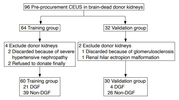
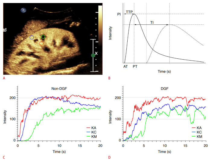
Methods: In total, 90 brain-dead donor kidneys (training group, n=60; validation group, n=30) examined between August 2020 and November 2022 were recruited in this prospective study. CEUS was performed on the kidneys of brain-dead donors 24 hours before organ procurement and time-intensity curves were constructed. The main measures were arrival time, time to peak, and peak intensity of the kidney segmental arteries, cortex, and medulla. Recipients were divided into DGF and non-DGF groups according to early post-transplant graft function. The area under the receiver operating characteristic curve (AUC) was used to assess diagnostic performance.
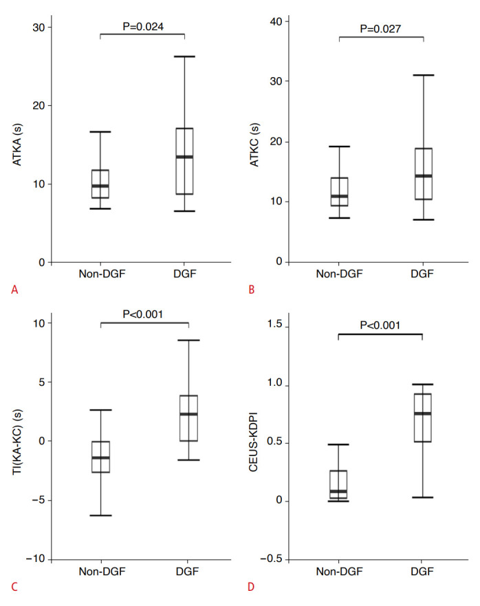
Results: The arrival time of the kidney segmental artery and cortex and the time interval between the time to peak of the segmental artery and cortex were identified as independent factors associated with DGF by multivariate stepwise regression analysis. A new index for the joint prediction model of three variables, the contrast-enhanced ultrasonography/Kidney Donor Profile index (CEUS-KDPI), was developed. CEUS-KDPI showed high accuracy for predicting DGF (training group: AUC, 0.91; sensitivity, 90.5%; specificity, 92.3%; validation group: AUC, 0.84; sensitivity, 75.0%; specificity, 92.3%).
Conclusion: CEUS-KDPI accurately predicted DGF after kidney transplantation. CEUS may be a potential noninvasive tool for bedside examinations before organ procurement and may be used to predict early renal function after kidney transplants kidneys from donors after brain death.
UltrasonographyMedicine-Radiology, Nuclear Medicine and Imaging
CiteScore
5.10
自引率
6.50%
发文量
78
审稿时长
15 weeks
期刊介绍:
Ultrasonography, the official English-language journal of the Korean Society of Ultrasound in Medicine (KSUM), is an international peer-reviewed academic journal dedicated to practice, research, technology, and education dealing with medical ultrasound. It is renamed from the Journal of Korean Society of Ultrasound in Medicine in January 2014, and published four times per year: January 1, April 1, July 1, and October 1. Original articles, technical notes, topical reviews, perspectives, pictorial essays, and timely editorial materials are published in Ultrasonography covering state-of-the-art content.
Ultrasonography aims to provide updated information on new diagnostic concepts and technical developments, including experimental animal studies using new equipment in addition to well-designed reviews of contemporary issues in patient care. Along with running KSUM Open, the annual international congress of KSUM, Ultrasonography also serves as a medium for cooperation among physicians and specialists from around the world who are focusing on various ultrasound technology and disease problems and relevant basic science.
 求助内容:
求助内容: 应助结果提醒方式:


