Aniruddha Mundhada , Sandhya Sundaram , Ramakrishnan Swaminathan , Lawrence D' Cruze , Satyavratan Govindarajan , Navaneethakrishna Makaram
{"title":"利用深度学习和可视化技术鉴别尿路上皮癌的组织病理学图像","authors":"Aniruddha Mundhada , Sandhya Sundaram , Ramakrishnan Swaminathan , Lawrence D' Cruze , Satyavratan Govindarajan , Navaneethakrishna Makaram","doi":"10.1016/j.jpi.2022.100155","DOIUrl":null,"url":null,"abstract":"<div><p>Artificial Intelligence is a tool poised to transform healthcare, with use in diagnostics and therapeutics. The widespread use of digital pathology has been due to the advent of whole slide imaging. Cheaper storage for digital images, along with unprecedented progress in artificial intelligence, have paved the synergy of these two fields. This has pushed the limits of traditional diagnosis using light microscopy, from a more subjective to a more objective method of looking at cases, incorporating grading too. The grading of histopathological images of urothelial carcinoma of the urinary bladder is important with direct implications for surgical management and prognosis.</p><p>In this study, the aim is to classify urothelial carcinoma into low and high grade based on the WHO 2016 classification. The hematoxylin and eosin-stained transurethral resection of bladder tumor (TURBT) samples of both low and high grade non-invasive papillary urothelial carcinoma were digitally scanned. Patches were extracted from these whole slide images to feed into a deep learning (Convolution Neural Network: CNN) model. Patches were segregated if they had tumor tissue and only included for model training if a threshold of 90% of tumor tissue per patch was seen. Various parameters of the deep learning model, known as hyperparameters, were optimized to get the best accuracy for grading or classification into low- and high-grade urothelial carcinoma. The model was robust with an overall accuracy of 90% after hyperparameter tuning. Visualization in the form of a class activation map using Grad-CAM was done. This indicates that such a model can be used as a companion diagnostic tool for grading of urothelial carcinoma. The probable causes of this accuracy are summarized along with the limitations of this study and future work possible.</p></div>","PeriodicalId":37769,"journal":{"name":"Journal of Pathology Informatics","volume":null,"pages":null},"PeriodicalIF":0.0000,"publicationDate":"2023-01-01","publicationTypes":"Journal Article","fieldsOfStudy":null,"isOpenAccess":false,"openAccessPdf":"https://ftp.ncbi.nlm.nih.gov/pub/pmc/oa_pdf/6c/f9/main.PMC9747506.pdf","citationCount":"3","resultStr":"{\"title\":\"Differentiation of urothelial carcinoma in histopathology images using deep learning and visualization\",\"authors\":\"Aniruddha Mundhada , Sandhya Sundaram , Ramakrishnan Swaminathan , Lawrence D' Cruze , Satyavratan Govindarajan , Navaneethakrishna Makaram\",\"doi\":\"10.1016/j.jpi.2022.100155\",\"DOIUrl\":null,\"url\":null,\"abstract\":\"<div><p>Artificial Intelligence is a tool poised to transform healthcare, with use in diagnostics and therapeutics. The widespread use of digital pathology has been due to the advent of whole slide imaging. Cheaper storage for digital images, along with unprecedented progress in artificial intelligence, have paved the synergy of these two fields. This has pushed the limits of traditional diagnosis using light microscopy, from a more subjective to a more objective method of looking at cases, incorporating grading too. The grading of histopathological images of urothelial carcinoma of the urinary bladder is important with direct implications for surgical management and prognosis.</p><p>In this study, the aim is to classify urothelial carcinoma into low and high grade based on the WHO 2016 classification. The hematoxylin and eosin-stained transurethral resection of bladder tumor (TURBT) samples of both low and high grade non-invasive papillary urothelial carcinoma were digitally scanned. Patches were extracted from these whole slide images to feed into a deep learning (Convolution Neural Network: CNN) model. Patches were segregated if they had tumor tissue and only included for model training if a threshold of 90% of tumor tissue per patch was seen. Various parameters of the deep learning model, known as hyperparameters, were optimized to get the best accuracy for grading or classification into low- and high-grade urothelial carcinoma. The model was robust with an overall accuracy of 90% after hyperparameter tuning. Visualization in the form of a class activation map using Grad-CAM was done. This indicates that such a model can be used as a companion diagnostic tool for grading of urothelial carcinoma. The probable causes of this accuracy are summarized along with the limitations of this study and future work possible.</p></div>\",\"PeriodicalId\":37769,\"journal\":{\"name\":\"Journal of Pathology Informatics\",\"volume\":null,\"pages\":null},\"PeriodicalIF\":0.0000,\"publicationDate\":\"2023-01-01\",\"publicationTypes\":\"Journal Article\",\"fieldsOfStudy\":null,\"isOpenAccess\":false,\"openAccessPdf\":\"https://ftp.ncbi.nlm.nih.gov/pub/pmc/oa_pdf/6c/f9/main.PMC9747506.pdf\",\"citationCount\":\"3\",\"resultStr\":null,\"platform\":\"Semanticscholar\",\"paperid\":null,\"PeriodicalName\":\"Journal of Pathology Informatics\",\"FirstCategoryId\":\"1085\",\"ListUrlMain\":\"https://www.sciencedirect.com/science/article/pii/S2153353922007490\",\"RegionNum\":0,\"RegionCategory\":null,\"ArticlePicture\":[],\"TitleCN\":null,\"AbstractTextCN\":null,\"PMCID\":null,\"EPubDate\":\"\",\"PubModel\":\"\",\"JCR\":\"Q2\",\"JCRName\":\"Medicine\",\"Score\":null,\"Total\":0}","platform":"Semanticscholar","paperid":null,"PeriodicalName":"Journal of Pathology Informatics","FirstCategoryId":"1085","ListUrlMain":"https://www.sciencedirect.com/science/article/pii/S2153353922007490","RegionNum":0,"RegionCategory":null,"ArticlePicture":[],"TitleCN":null,"AbstractTextCN":null,"PMCID":null,"EPubDate":"","PubModel":"","JCR":"Q2","JCRName":"Medicine","Score":null,"Total":0}
Differentiation of urothelial carcinoma in histopathology images using deep learning and visualization
Artificial Intelligence is a tool poised to transform healthcare, with use in diagnostics and therapeutics. The widespread use of digital pathology has been due to the advent of whole slide imaging. Cheaper storage for digital images, along with unprecedented progress in artificial intelligence, have paved the synergy of these two fields. This has pushed the limits of traditional diagnosis using light microscopy, from a more subjective to a more objective method of looking at cases, incorporating grading too. The grading of histopathological images of urothelial carcinoma of the urinary bladder is important with direct implications for surgical management and prognosis.
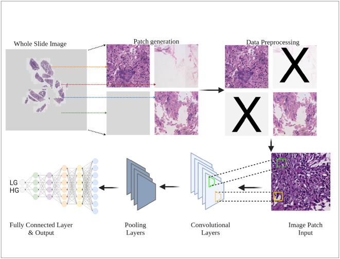
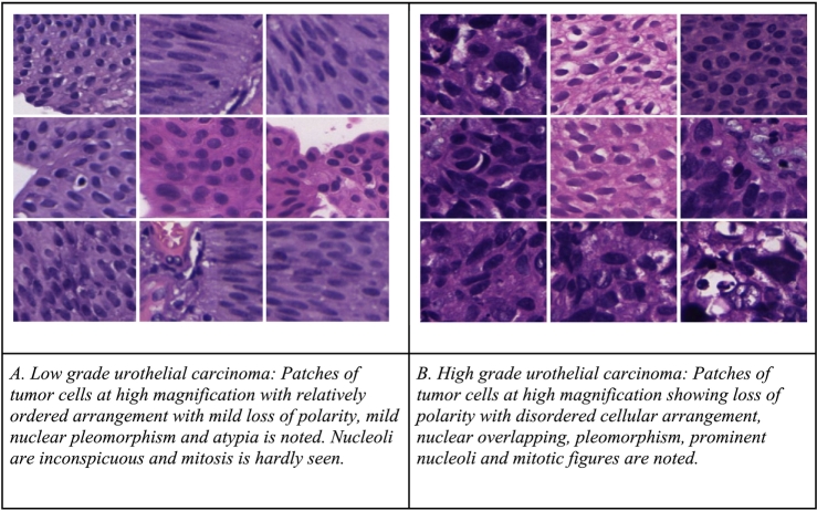
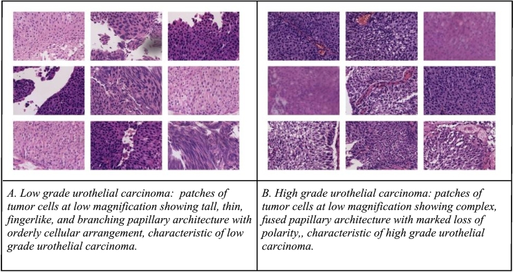
In this study, the aim is to classify urothelial carcinoma into low and high grade based on the WHO 2016 classification. The hematoxylin and eosin-stained transurethral resection of bladder tumor (TURBT) samples of both low and high grade non-invasive papillary urothelial carcinoma were digitally scanned. Patches were extracted from these whole slide images to feed into a deep learning (Convolution Neural Network: CNN) model. Patches were segregated if they had tumor tissue and only included for model training if a threshold of 90% of tumor tissue per patch was seen. Various parameters of the deep learning model, known as hyperparameters, were optimized to get the best accuracy for grading or classification into low- and high-grade urothelial carcinoma. The model was robust with an overall accuracy of 90% after hyperparameter tuning. Visualization in the form of a class activation map using Grad-CAM was done. This indicates that such a model can be used as a companion diagnostic tool for grading of urothelial carcinoma. The probable causes of this accuracy are summarized along with the limitations of this study and future work possible.
期刊介绍:
The Journal of Pathology Informatics (JPI) is an open access peer-reviewed journal dedicated to the advancement of pathology informatics. This is the official journal of the Association for Pathology Informatics (API). The journal aims to publish broadly about pathology informatics and freely disseminate all articles worldwide. This journal is of interest to pathologists, informaticians, academics, researchers, health IT specialists, information officers, IT staff, vendors, and anyone with an interest in informatics. We encourage submissions from anyone with an interest in the field of pathology informatics. We publish all types of papers related to pathology informatics including original research articles, technical notes, reviews, viewpoints, commentaries, editorials, symposia, meeting abstracts, book reviews, and correspondence to the editors. All submissions are subject to rigorous peer review by the well-regarded editorial board and by expert referees in appropriate specialties.

 求助内容:
求助内容: 应助结果提醒方式:


