Jung Hee Byon, Gong Yong Jin, Young Min Han, Eun Jung Choi, Kum Ju Chae, Eun Hae Park
{"title":"胸部CT正常患者吸烟习惯与慢性阻塞性肺疾病的定量CT分析","authors":"Jung Hee Byon, Gong Yong Jin, Young Min Han, Eun Jung Choi, Kum Ju Chae, Eun Hae Park","doi":"10.3348/jksr.2022.0130","DOIUrl":null,"url":null,"abstract":"<p><strong>Purpose: </strong>To assess normal CT scans with quantitative CT (QCT) analysis based on smoking habits and chronic obstructive pulmonary disease (COPD).</p><p><strong>Materials and methods: </strong>From January 2013 to December 2014, 90 male patients with normal chest CT and quantification analysis results were enrolled in our study [non-COPD never-smokers (<i>n</i> = 38) and smokers (<i>n</i> = 45), COPD smokers (<i>n</i> = 7)]. In addition, an age-matched cohort study was performed for seven smokers with COPD. The square root of the wall area of a hypothetical bronchus of internal perimeter 10 mm (Pi10), skewness, kurtosis, mean lung attenuation (MLA), and percentage of low attenuation area (%LAA) were evaluated.</p><p><strong>Results: </strong>Among patients without COPD, the Pi10 of smokers (4.176 ± 0.282) was about 0.1 mm thicker than that of never-smokers (4.070 ± 0.191, <i>p</i> = 0.047), and skewness and kurtosis of smokers (2.628 ± 0.484 and 6.448 ± 3.427) were lower than never-smokers (2.884 ± 0.624, <i>p</i> = 0.038 and 8.594 ± 4.944, <i>p</i> = 0.02). The Pi10 of COPD smokers (4.429 ± 0.435, <i>n</i> = 7) was about 0.4 mm thicker than never-smokers without COPD (3.996 ± 0.115, <i>n</i> = 14, <i>p</i> = 0.005). There were no significant differences in MLA and %LAA between groups (<i>p</i> > 0.05).</p><p><strong>Conclusion: </strong>Even on normal CT scans, QCT showed that the airway walls of smokers are thicker than never-smokers regardless of COPD and it preceded lung parenchymal changes.</p>","PeriodicalId":17455,"journal":{"name":"Journal of the Korean Society of Radiology","volume":"84 4","pages":"900-910"},"PeriodicalIF":0.0000,"publicationDate":"2023-07-01","publicationTypes":"Journal Article","fieldsOfStudy":null,"isOpenAccess":false,"openAccessPdf":"https://ftp.ncbi.nlm.nih.gov/pub/pmc/oa_pdf/96/b7/jksr-84-900.PMC10407071.pdf","citationCount":"0","resultStr":"{\"title\":\"Quantitative CT Analysis Based on Smoking Habits and Chronic Obstructive Pulmonary Disease in Patients with Normal Chest CT.\",\"authors\":\"Jung Hee Byon, Gong Yong Jin, Young Min Han, Eun Jung Choi, Kum Ju Chae, Eun Hae Park\",\"doi\":\"10.3348/jksr.2022.0130\",\"DOIUrl\":null,\"url\":null,\"abstract\":\"<p><strong>Purpose: </strong>To assess normal CT scans with quantitative CT (QCT) analysis based on smoking habits and chronic obstructive pulmonary disease (COPD).</p><p><strong>Materials and methods: </strong>From January 2013 to December 2014, 90 male patients with normal chest CT and quantification analysis results were enrolled in our study [non-COPD never-smokers (<i>n</i> = 38) and smokers (<i>n</i> = 45), COPD smokers (<i>n</i> = 7)]. In addition, an age-matched cohort study was performed for seven smokers with COPD. The square root of the wall area of a hypothetical bronchus of internal perimeter 10 mm (Pi10), skewness, kurtosis, mean lung attenuation (MLA), and percentage of low attenuation area (%LAA) were evaluated.</p><p><strong>Results: </strong>Among patients without COPD, the Pi10 of smokers (4.176 ± 0.282) was about 0.1 mm thicker than that of never-smokers (4.070 ± 0.191, <i>p</i> = 0.047), and skewness and kurtosis of smokers (2.628 ± 0.484 and 6.448 ± 3.427) were lower than never-smokers (2.884 ± 0.624, <i>p</i> = 0.038 and 8.594 ± 4.944, <i>p</i> = 0.02). The Pi10 of COPD smokers (4.429 ± 0.435, <i>n</i> = 7) was about 0.4 mm thicker than never-smokers without COPD (3.996 ± 0.115, <i>n</i> = 14, <i>p</i> = 0.005). There were no significant differences in MLA and %LAA between groups (<i>p</i> > 0.05).</p><p><strong>Conclusion: </strong>Even on normal CT scans, QCT showed that the airway walls of smokers are thicker than never-smokers regardless of COPD and it preceded lung parenchymal changes.</p>\",\"PeriodicalId\":17455,\"journal\":{\"name\":\"Journal of the Korean Society of Radiology\",\"volume\":\"84 4\",\"pages\":\"900-910\"},\"PeriodicalIF\":0.0000,\"publicationDate\":\"2023-07-01\",\"publicationTypes\":\"Journal Article\",\"fieldsOfStudy\":null,\"isOpenAccess\":false,\"openAccessPdf\":\"https://ftp.ncbi.nlm.nih.gov/pub/pmc/oa_pdf/96/b7/jksr-84-900.PMC10407071.pdf\",\"citationCount\":\"0\",\"resultStr\":null,\"platform\":\"Semanticscholar\",\"paperid\":null,\"PeriodicalName\":\"Journal of the Korean Society of Radiology\",\"FirstCategoryId\":\"1085\",\"ListUrlMain\":\"https://doi.org/10.3348/jksr.2022.0130\",\"RegionNum\":0,\"RegionCategory\":null,\"ArticlePicture\":[],\"TitleCN\":null,\"AbstractTextCN\":null,\"PMCID\":null,\"EPubDate\":\"\",\"PubModel\":\"\",\"JCR\":\"Q4\",\"JCRName\":\"Medicine\",\"Score\":null,\"Total\":0}","platform":"Semanticscholar","paperid":null,"PeriodicalName":"Journal of the Korean Society of Radiology","FirstCategoryId":"1085","ListUrlMain":"https://doi.org/10.3348/jksr.2022.0130","RegionNum":0,"RegionCategory":null,"ArticlePicture":[],"TitleCN":null,"AbstractTextCN":null,"PMCID":null,"EPubDate":"","PubModel":"","JCR":"Q4","JCRName":"Medicine","Score":null,"Total":0}
Quantitative CT Analysis Based on Smoking Habits and Chronic Obstructive Pulmonary Disease in Patients with Normal Chest CT.
Purpose: To assess normal CT scans with quantitative CT (QCT) analysis based on smoking habits and chronic obstructive pulmonary disease (COPD).
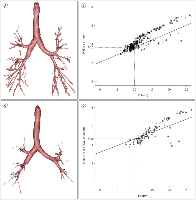
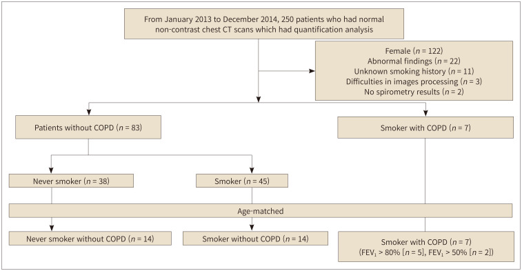
Materials and methods: From January 2013 to December 2014, 90 male patients with normal chest CT and quantification analysis results were enrolled in our study [non-COPD never-smokers (n = 38) and smokers (n = 45), COPD smokers (n = 7)]. In addition, an age-matched cohort study was performed for seven smokers with COPD. The square root of the wall area of a hypothetical bronchus of internal perimeter 10 mm (Pi10), skewness, kurtosis, mean lung attenuation (MLA), and percentage of low attenuation area (%LAA) were evaluated.
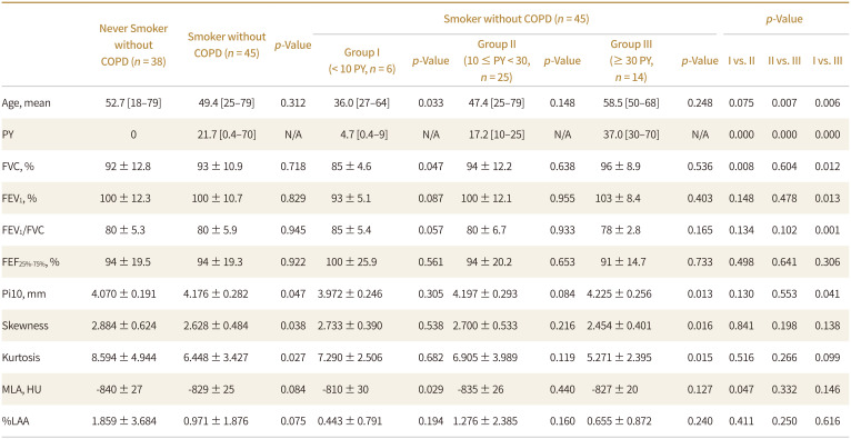
Results: Among patients without COPD, the Pi10 of smokers (4.176 ± 0.282) was about 0.1 mm thicker than that of never-smokers (4.070 ± 0.191, p = 0.047), and skewness and kurtosis of smokers (2.628 ± 0.484 and 6.448 ± 3.427) were lower than never-smokers (2.884 ± 0.624, p = 0.038 and 8.594 ± 4.944, p = 0.02). The Pi10 of COPD smokers (4.429 ± 0.435, n = 7) was about 0.4 mm thicker than never-smokers without COPD (3.996 ± 0.115, n = 14, p = 0.005). There were no significant differences in MLA and %LAA between groups (p > 0.05).
Conclusion: Even on normal CT scans, QCT showed that the airway walls of smokers are thicker than never-smokers regardless of COPD and it preceded lung parenchymal changes.

 求助内容:
求助内容: 应助结果提醒方式:


