小眼症的动物和细胞模型。
摘要
小眼症是一种罕见的发育性眼病,每7000名新生儿中就有1名患有这种疾病。它被定义为一个小的(轴长⩾2标准差低于年龄调整平均值)发育不全的眼睛,由妊娠早期的遗传或环境因素破坏眼睛发育引起。临床表型异质性存在于不同严重程度以及相关眼部和全身特征的患者中。据报道,多达11%的失明儿童患有小眼症,但目前尚无治疗方法。通过确定小眼的病因并了解眼睛发育的机制是如何被破坏的,我们可以更好地了解发病机制。动物模型,主要是小鼠、斑马鱼和非洲爪蟾,已经提供了关于眼睛发育的基因调控以及这些途径的干扰如何导致小眼症的广泛信息。然而,物种之间存在差异,因此细胞模型,如患者衍生的诱导多能干细胞(iPSC)视小泡,现在正被用于对人类疾病过程提供更深入的了解。3D细胞建模技术的进步增强了研究人员研究眼睛发育过程中不同细胞类型相互作用的能力。通过提高对小眼症的分子知识,可以开发预防性或产后治疗,同时建立基因型-表型相关性,为患者提供适当的预后、多学科护理和知情的遗传咨询。这篇综述总结了小眼球动物和细胞模型的一些关键发现,并讨论了如何在未来使用创新的新模型来加深我们的理解。简明语言总结:眼部疾病的动物和细胞模型,小眼症(小眼)小眼症是一种罕见的儿童先天性疾病,意思是眼睛小而发育不全。前3年环境的遗传变化或变异 怀孕数月会破坏眼睛的早期发育,导致小眼症。高达11%的失明儿童患有小眼症,但目前尚无治疗方法。通过了解眼睛发育所需的基因,我们可以确定基因变化或环境因素的破坏是如何导致这种情况的。这有助于我们了解小眼症发生的原因,并确保为患者提供适当的临床护理和遗传咨询建议。此外,通过了解小眼症的原因,研究人员可以开发预防或减轻这种情况严重程度的治疗方法。动物模型,特别是小鼠、斑马鱼和青蛙,由于同样的基因/环境变化,它们也会长出小眼睛,帮助我们了解了对眼睛发育很重要的基因,这些基因在被破坏时会导致出生时的眼睛缺陷。研究患者在实验室中生长的细胞可以进一步帮助研究人员了解基因的变化如何影响其功能。动物和细胞模型都可以用于开发和测试新药,为小眼症患者提供治疗选择。这篇综述总结了小眼球动物和细胞模型的关键发现,并讨论了如何在未来使用创新的新模型来加深我们的理解。
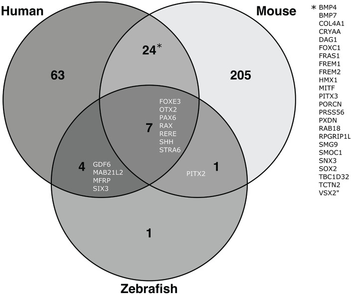

Microphthalmia is a rare developmental eye disorder affecting 1 in 7000 births. It is defined as a small (axial length ⩾2 standard deviations below the age-adjusted mean) underdeveloped eye, caused by disruption of ocular development through genetic or environmental factors in the first trimester of pregnancy. Clinical phenotypic heterogeneity exists amongst patients with varying levels of severity, and associated ocular and systemic features. Up to 11% of blind children are reported to have microphthalmia, yet currently no treatments are available. By identifying the aetiology of microphthalmia and understanding how the mechanisms of eye development are disrupted, we can gain a better understanding of the pathogenesis. Animal models, mainly mouse, zebrafish and Xenopus, have provided extensive information on the genetic regulation of oculogenesis, and how perturbation of these pathways leads to microphthalmia. However, differences exist between species, hence cellular models, such as patient-derived induced pluripotent stem cell (iPSC) optic vesicles, are now being used to provide greater insights into the human disease process. Progress in 3D cellular modelling techniques has enhanced the ability of researchers to study interactions of different cell types during eye development. Through improved molecular knowledge of microphthalmia, preventative or postnatal therapies may be developed, together with establishing genotype-phenotype correlations in order to provide patients with the appropriate prognosis, multidisciplinary care and informed genetic counselling. This review summarises some key discoveries from animal and cellular models of microphthalmia and discusses how innovative new models can be used to further our understanding in the future.
Plain language summary: Animal and Cellular Models of the Eye Disorder, Microphthalmia (Small Eye) Microphthalmia, meaning a small, underdeveloped eye, is a rare disorder that children are born with. Genetic changes or variations in the environment during the first 3 months of pregnancy can disrupt early development of the eye, resulting in microphthalmia. Up to 11% of blind children have microphthalmia, yet currently no treatments are available. By understanding the genes necessary for eye development, we can determine how disruption by genetic changes or environmental factors can cause this condition. This helps us understand why microphthalmia occurs, and ensure patients are provided with the appropriate clinical care and genetic counselling advice. Additionally, by understanding the causes of microphthalmia, researchers can develop treatments to prevent or reduce the severity of this condition. Animal models, particularly mice, zebrafish and frogs, which can also develop small eyes due to the same genetic/environmental changes, have helped us understand the genes which are important for eye development and can cause birth eye defects when disrupted. Studying a patient's own cells grown in the laboratory can further help researchers understand how changes in genes affect their function. Both animal and cellular models can be used to develop and test new drugs, which could provide treatment options for patients living with microphthalmia. This review summarises the key discoveries from animal and cellular models of microphthalmia and discusses how innovative new models can be used to further our understanding in the future.

 求助内容:
求助内容: 应助结果提醒方式:


