{"title":"三维T1-Turbo自旋回波成像对颅内支架置入术评价的价值。","authors":"Hiroyuki Mizuno, Masanori Aihara, Koji Sato, Chikashi Negishi, Nobuo Sasaguchi, Hideyuki Kurihara, Yuhei Yoshimoto","doi":"10.5797/jnet.oa.2022-0039","DOIUrl":null,"url":null,"abstract":"<p><strong>Objective: </strong>Evaluation of intracranial stent placement by MRI suffers the problems of signal artifacts during time-of-flight MRA (TOF-MRA). Therefore, angiographic examination is required for detailed intravascular assessment of the stent placement site. Recently, 3D T1-turbo spin echo (3D-TSE) has been developed for evaluation of carotid artery stent placement. We investigated the use of the 3D-TSE imaging method for the evaluation of intracranial vascular stent placement.</p><p><strong>Methods: </strong>The subjects consisted of nine patients who underwent intracranial vascular stent placement between April 2015 and December 2019. Postoperatively, the lumens of the placed stents were measured by TOF-MRA, DSA, and 3D-TSE imaging. Analysis was performed by type of stent and placement site.</p><p><strong>Results: </strong>The stents used were Neuroform Atlas (3 patients), LVIS (3 patients), LVIS Jr (2 patients), and Integrity (1 patient). TOF-MRA of the stent placement site showed defects in the image or poor visualization in all nine patients, whereas 3D-TSE imaging visualized the lumen at the stent indwelling site in all patients. The blood vessel diameter measured by the DSA and 3D-TSE imaging exhibited positive correlations regardless of the stent type and placement site.</p><p><strong>Conclusion: </strong>3D-TSE imaging allows visualization of the lumen of the site of an intracranial vascular stent, regardless of the type of stent or the vessel. Thus, this method may be useful for evaluating the vascular lumen of a lesion.</p>","PeriodicalId":73856,"journal":{"name":"Journal of neuroendovascular therapy","volume":"17 1","pages":"1-7"},"PeriodicalIF":0.0000,"publicationDate":"2023-01-01","publicationTypes":"Journal Article","fieldsOfStudy":null,"isOpenAccess":false,"openAccessPdf":"https://ftp.ncbi.nlm.nih.gov/pub/pmc/oa_pdf/32/28/jnet-17-01.PMC10370516.pdf","citationCount":"0","resultStr":"{\"title\":\"Usefulness of 3D T1-Turbo Spin Echo Imaging for the Evaluation of Intracranial Stent Placement.\",\"authors\":\"Hiroyuki Mizuno, Masanori Aihara, Koji Sato, Chikashi Negishi, Nobuo Sasaguchi, Hideyuki Kurihara, Yuhei Yoshimoto\",\"doi\":\"10.5797/jnet.oa.2022-0039\",\"DOIUrl\":null,\"url\":null,\"abstract\":\"<p><strong>Objective: </strong>Evaluation of intracranial stent placement by MRI suffers the problems of signal artifacts during time-of-flight MRA (TOF-MRA). Therefore, angiographic examination is required for detailed intravascular assessment of the stent placement site. Recently, 3D T1-turbo spin echo (3D-TSE) has been developed for evaluation of carotid artery stent placement. We investigated the use of the 3D-TSE imaging method for the evaluation of intracranial vascular stent placement.</p><p><strong>Methods: </strong>The subjects consisted of nine patients who underwent intracranial vascular stent placement between April 2015 and December 2019. Postoperatively, the lumens of the placed stents were measured by TOF-MRA, DSA, and 3D-TSE imaging. Analysis was performed by type of stent and placement site.</p><p><strong>Results: </strong>The stents used were Neuroform Atlas (3 patients), LVIS (3 patients), LVIS Jr (2 patients), and Integrity (1 patient). TOF-MRA of the stent placement site showed defects in the image or poor visualization in all nine patients, whereas 3D-TSE imaging visualized the lumen at the stent indwelling site in all patients. The blood vessel diameter measured by the DSA and 3D-TSE imaging exhibited positive correlations regardless of the stent type and placement site.</p><p><strong>Conclusion: </strong>3D-TSE imaging allows visualization of the lumen of the site of an intracranial vascular stent, regardless of the type of stent or the vessel. Thus, this method may be useful for evaluating the vascular lumen of a lesion.</p>\",\"PeriodicalId\":73856,\"journal\":{\"name\":\"Journal of neuroendovascular therapy\",\"volume\":\"17 1\",\"pages\":\"1-7\"},\"PeriodicalIF\":0.0000,\"publicationDate\":\"2023-01-01\",\"publicationTypes\":\"Journal Article\",\"fieldsOfStudy\":null,\"isOpenAccess\":false,\"openAccessPdf\":\"https://ftp.ncbi.nlm.nih.gov/pub/pmc/oa_pdf/32/28/jnet-17-01.PMC10370516.pdf\",\"citationCount\":\"0\",\"resultStr\":null,\"platform\":\"Semanticscholar\",\"paperid\":null,\"PeriodicalName\":\"Journal of neuroendovascular therapy\",\"FirstCategoryId\":\"1085\",\"ListUrlMain\":\"https://doi.org/10.5797/jnet.oa.2022-0039\",\"RegionNum\":0,\"RegionCategory\":null,\"ArticlePicture\":[],\"TitleCN\":null,\"AbstractTextCN\":null,\"PMCID\":null,\"EPubDate\":\"\",\"PubModel\":\"\",\"JCR\":\"\",\"JCRName\":\"\",\"Score\":null,\"Total\":0}","platform":"Semanticscholar","paperid":null,"PeriodicalName":"Journal of neuroendovascular therapy","FirstCategoryId":"1085","ListUrlMain":"https://doi.org/10.5797/jnet.oa.2022-0039","RegionNum":0,"RegionCategory":null,"ArticlePicture":[],"TitleCN":null,"AbstractTextCN":null,"PMCID":null,"EPubDate":"","PubModel":"","JCR":"","JCRName":"","Score":null,"Total":0}
Usefulness of 3D T1-Turbo Spin Echo Imaging for the Evaluation of Intracranial Stent Placement.
Objective: Evaluation of intracranial stent placement by MRI suffers the problems of signal artifacts during time-of-flight MRA (TOF-MRA). Therefore, angiographic examination is required for detailed intravascular assessment of the stent placement site. Recently, 3D T1-turbo spin echo (3D-TSE) has been developed for evaluation of carotid artery stent placement. We investigated the use of the 3D-TSE imaging method for the evaluation of intracranial vascular stent placement.
Methods: The subjects consisted of nine patients who underwent intracranial vascular stent placement between April 2015 and December 2019. Postoperatively, the lumens of the placed stents were measured by TOF-MRA, DSA, and 3D-TSE imaging. Analysis was performed by type of stent and placement site.
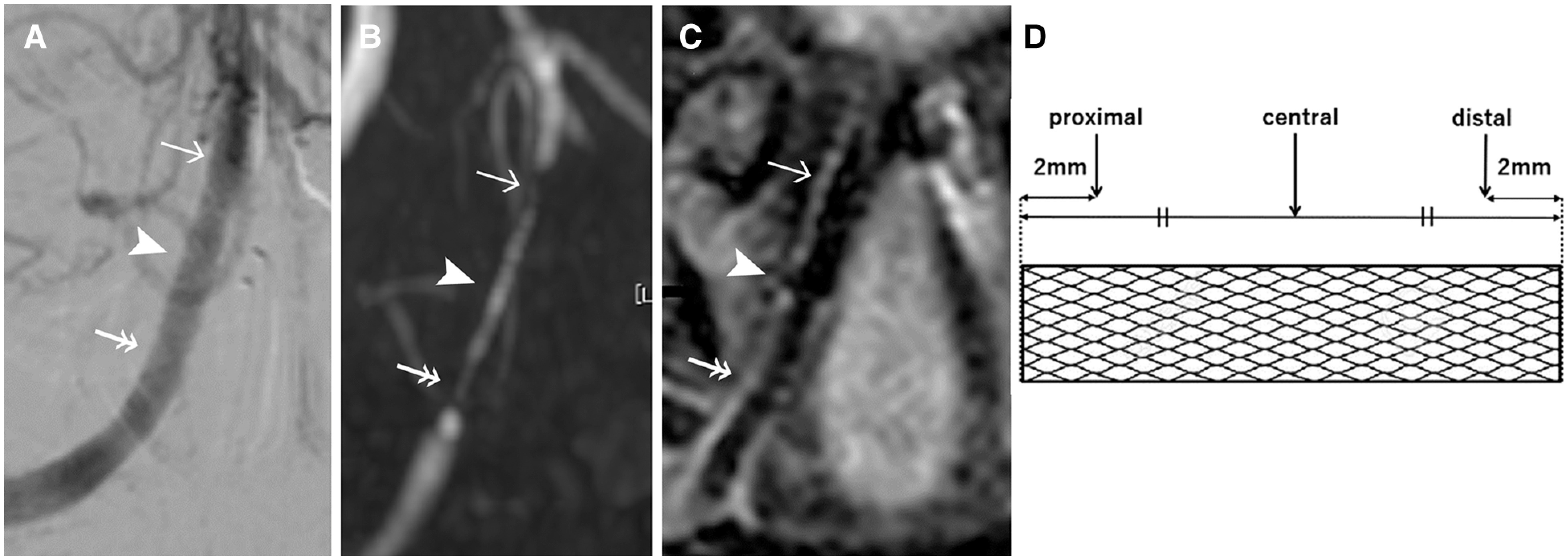
Results: The stents used were Neuroform Atlas (3 patients), LVIS (3 patients), LVIS Jr (2 patients), and Integrity (1 patient). TOF-MRA of the stent placement site showed defects in the image or poor visualization in all nine patients, whereas 3D-TSE imaging visualized the lumen at the stent indwelling site in all patients. The blood vessel diameter measured by the DSA and 3D-TSE imaging exhibited positive correlations regardless of the stent type and placement site.
Conclusion: 3D-TSE imaging allows visualization of the lumen of the site of an intracranial vascular stent, regardless of the type of stent or the vessel. Thus, this method may be useful for evaluating the vascular lumen of a lesion.

 求助内容:
求助内容: 应助结果提醒方式:


