{"title":"正常胸腺及其随年龄变化的多检波器计算机断层扫描评价。","authors":"Suprava Naik, Aishvarya Shri, Simran Sidhu, Yuvraj Lahre, Nerbadyswari Deep Bag, Sanjeev Kumar Bhoi, Sudipta Mohakud","doi":"10.4103/jmas.jmas_25_23","DOIUrl":null,"url":null,"abstract":"<p><strong>Background: </strong>Thymus is a T-cell-producing lymphoid organ that appears prominent in the paediatric population and involutes in size with ageing. The gland shows a wide variety of appearances across different age groups. The purpose of the study is to evaluate the computed tomography (CT) appearance of thymus gland in the normal population with a focus on size, CT attenuation and fatty infiltration in different age groups.</p><p><strong>Patients and methods: </strong>This is a retrospective study done after taking approval from the Institutional Ethics Committee. Patients undergone CT scans of the thorax were identified from our database. All evaluations were done in non-contrast CT scans. Patients having underlying diseases that may have associated thymic abnormality were excluded. The appearance of thymus and the presence of fatty replacement were assessed. The size of thymus (length and thickness of right limb and left limb) and non-contrast CT Hounsfield unit (HU) value of thymic tissue were measured and compared in various age groups.</p><p><strong>Results: </strong>Four hundred and fifty patients were included, 262 (58.2%) were male. Mean age was 33.6 ± 17.1 years, range (3 months-80 years). The size of thymus was observed to decrease with increasing age. The mean age of complete fatty replacement in our study was 45 years. Complete fatty replacement was noted in all cases with an age of more than 60 years. The most common shape was arrowhead, and the most common location was pre-aortic and para-aortic location. Non-contrast CT HU value was maximum in infants and gradually decreased with advancing age.</p><p><strong>Conclusion: </strong>Even normal thymus can show varied appearance on CT which changes with the age of the patient being imaged. A comparison with normative data could help differentiate normal from abnormal glands to avoid unnecessary intervention.</p>","PeriodicalId":48905,"journal":{"name":"Journal of Minimal Access Surgery","volume":" ","pages":"101-106"},"PeriodicalIF":1.1000,"publicationDate":"2025-04-01","publicationTypes":"Journal Article","fieldsOfStudy":null,"isOpenAccess":false,"openAccessPdf":"https://www.ncbi.nlm.nih.gov/pmc/articles/PMC12054957/pdf/","citationCount":"0","resultStr":"{\"title\":\"Multidetector computed tomography evaluation of normal thymus and variations with age.\",\"authors\":\"Suprava Naik, Aishvarya Shri, Simran Sidhu, Yuvraj Lahre, Nerbadyswari Deep Bag, Sanjeev Kumar Bhoi, Sudipta Mohakud\",\"doi\":\"10.4103/jmas.jmas_25_23\",\"DOIUrl\":null,\"url\":null,\"abstract\":\"<p><strong>Background: </strong>Thymus is a T-cell-producing lymphoid organ that appears prominent in the paediatric population and involutes in size with ageing. The gland shows a wide variety of appearances across different age groups. The purpose of the study is to evaluate the computed tomography (CT) appearance of thymus gland in the normal population with a focus on size, CT attenuation and fatty infiltration in different age groups.</p><p><strong>Patients and methods: </strong>This is a retrospective study done after taking approval from the Institutional Ethics Committee. Patients undergone CT scans of the thorax were identified from our database. All evaluations were done in non-contrast CT scans. Patients having underlying diseases that may have associated thymic abnormality were excluded. The appearance of thymus and the presence of fatty replacement were assessed. The size of thymus (length and thickness of right limb and left limb) and non-contrast CT Hounsfield unit (HU) value of thymic tissue were measured and compared in various age groups.</p><p><strong>Results: </strong>Four hundred and fifty patients were included, 262 (58.2%) were male. Mean age was 33.6 ± 17.1 years, range (3 months-80 years). The size of thymus was observed to decrease with increasing age. The mean age of complete fatty replacement in our study was 45 years. Complete fatty replacement was noted in all cases with an age of more than 60 years. The most common shape was arrowhead, and the most common location was pre-aortic and para-aortic location. Non-contrast CT HU value was maximum in infants and gradually decreased with advancing age.</p><p><strong>Conclusion: </strong>Even normal thymus can show varied appearance on CT which changes with the age of the patient being imaged. A comparison with normative data could help differentiate normal from abnormal glands to avoid unnecessary intervention.</p>\",\"PeriodicalId\":48905,\"journal\":{\"name\":\"Journal of Minimal Access Surgery\",\"volume\":\" \",\"pages\":\"101-106\"},\"PeriodicalIF\":1.1000,\"publicationDate\":\"2025-04-01\",\"publicationTypes\":\"Journal Article\",\"fieldsOfStudy\":null,\"isOpenAccess\":false,\"openAccessPdf\":\"https://www.ncbi.nlm.nih.gov/pmc/articles/PMC12054957/pdf/\",\"citationCount\":\"0\",\"resultStr\":null,\"platform\":\"Semanticscholar\",\"paperid\":null,\"PeriodicalName\":\"Journal of Minimal Access Surgery\",\"FirstCategoryId\":\"3\",\"ListUrlMain\":\"https://doi.org/10.4103/jmas.jmas_25_23\",\"RegionNum\":4,\"RegionCategory\":\"医学\",\"ArticlePicture\":[],\"TitleCN\":null,\"AbstractTextCN\":null,\"PMCID\":null,\"EPubDate\":\"2023/7/5 0:00:00\",\"PubModel\":\"Epub\",\"JCR\":\"Q3\",\"JCRName\":\"SURGERY\",\"Score\":null,\"Total\":0}","platform":"Semanticscholar","paperid":null,"PeriodicalName":"Journal of Minimal Access Surgery","FirstCategoryId":"3","ListUrlMain":"https://doi.org/10.4103/jmas.jmas_25_23","RegionNum":4,"RegionCategory":"医学","ArticlePicture":[],"TitleCN":null,"AbstractTextCN":null,"PMCID":null,"EPubDate":"2023/7/5 0:00:00","PubModel":"Epub","JCR":"Q3","JCRName":"SURGERY","Score":null,"Total":0}
Multidetector computed tomography evaluation of normal thymus and variations with age.
Background: Thymus is a T-cell-producing lymphoid organ that appears prominent in the paediatric population and involutes in size with ageing. The gland shows a wide variety of appearances across different age groups. The purpose of the study is to evaluate the computed tomography (CT) appearance of thymus gland in the normal population with a focus on size, CT attenuation and fatty infiltration in different age groups.
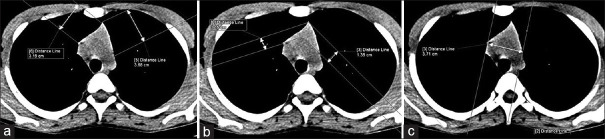
Patients and methods: This is a retrospective study done after taking approval from the Institutional Ethics Committee. Patients undergone CT scans of the thorax were identified from our database. All evaluations were done in non-contrast CT scans. Patients having underlying diseases that may have associated thymic abnormality were excluded. The appearance of thymus and the presence of fatty replacement were assessed. The size of thymus (length and thickness of right limb and left limb) and non-contrast CT Hounsfield unit (HU) value of thymic tissue were measured and compared in various age groups.
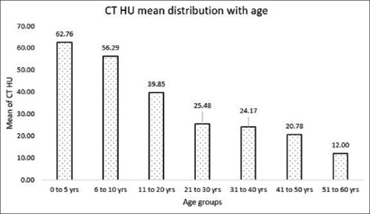
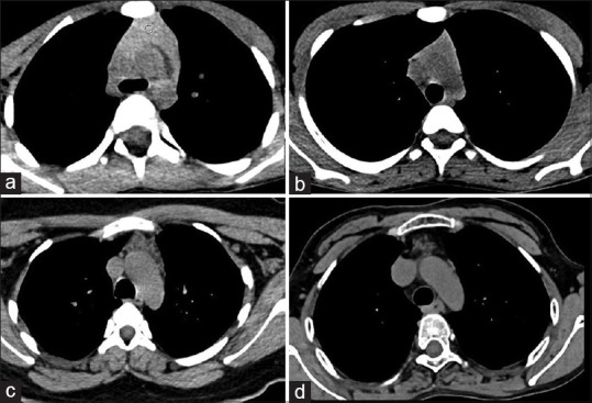
Results: Four hundred and fifty patients were included, 262 (58.2%) were male. Mean age was 33.6 ± 17.1 years, range (3 months-80 years). The size of thymus was observed to decrease with increasing age. The mean age of complete fatty replacement in our study was 45 years. Complete fatty replacement was noted in all cases with an age of more than 60 years. The most common shape was arrowhead, and the most common location was pre-aortic and para-aortic location. Non-contrast CT HU value was maximum in infants and gradually decreased with advancing age.
Conclusion: Even normal thymus can show varied appearance on CT which changes with the age of the patient being imaged. A comparison with normative data could help differentiate normal from abnormal glands to avoid unnecessary intervention.
期刊介绍:
Journal of Minimal Access Surgery (JMAS), the official publication of Indian Association of Gastrointestinal Endo Surgeons, launched in early 2005. The JMAS, a quarterly publication, is the first English-language journal from India, as also from this part of the world, dedicated to Minimal Access Surgery. The JMAS boasts an outstanding editorial board comprising of Indian and international experts in the field.
 求助内容:
求助内容: 应助结果提醒方式:


