Kai M Bracey, Margret Fye, Alisa Cario, Kung-Hsien Ho, Pi'illani Noguchi, Guoqiang Gu, Irina Kaverina
{"title":"葡萄糖刺激的KIF5B驱动的微管滑动在胰腺β细胞中组织微管网络。","authors":"Kai M Bracey, Margret Fye, Alisa Cario, Kung-Hsien Ho, Pi'illani Noguchi, Guoqiang Gu, Irina Kaverina","doi":"10.1101/2023.06.25.546468","DOIUrl":null,"url":null,"abstract":"<p><p>In pancreatic islet β cells, molecular motors use cytoskeletal polymers microtubules as tracks for intracellular transport of insulin secretory granules. The β-cell microtubule network has a complex architecture and is non-directional, which provides insulin granules at the cell periphery for rapid secretion response, yet to avoid over-secretion and subsequent hypoglycemia. We have previously characterized a peripheral sub-membrane microtubule array, which is critical for the withdrawal of excessive insulin granules from the secretion sites. Microtubules in β cells originate at the Golgi in the cell interior, and how the peripheral array is formed is unknown. Using real-time imaging and photo-kinetics approaches in clonal mouse pancreatic β cells MIN6, we now demonstrate that kinesin KIF5B, a motor protein with a capacity to transport microtubules as cargos, slides existing microtubules to the cell periphery and aligns them to each other along the plasma membrane. Moreover, like many physiological β-cell features, microtubule sliding is facilitated by a high glucose stimulus. These new data, together with our previous report that in high glucose sub-membrane MT array is destabilized to allow for robust secretion, indicate that MT sliding is another integral part of glucose-triggered microtubule remodeling, likely replacing destabilized peripheral microtubules to prevent their loss over time and β-cell malfunction.</p>","PeriodicalId":72407,"journal":{"name":"bioRxiv : the preprint server for biology","volume":" ","pages":""},"PeriodicalIF":0.0000,"publicationDate":"2025-05-05","publicationTypes":"Journal Article","fieldsOfStudy":null,"isOpenAccess":false,"openAccessPdf":"https://ftp.ncbi.nlm.nih.gov/pub/pmc/oa_pdf/ae/fa/nihpp-2023.06.25.546468v1.PMC10327020.pdf","citationCount":"0","resultStr":"{\"title\":\"Glucose-stimulated KIF5B-driven microtubule sliding organizes microtubule networks in pancreatic β cells.\",\"authors\":\"Kai M Bracey, Margret Fye, Alisa Cario, Kung-Hsien Ho, Pi'illani Noguchi, Guoqiang Gu, Irina Kaverina\",\"doi\":\"10.1101/2023.06.25.546468\",\"DOIUrl\":null,\"url\":null,\"abstract\":\"<p><p>In pancreatic islet β cells, molecular motors use cytoskeletal polymers microtubules as tracks for intracellular transport of insulin secretory granules. The β-cell microtubule network has a complex architecture and is non-directional, which provides insulin granules at the cell periphery for rapid secretion response, yet to avoid over-secretion and subsequent hypoglycemia. We have previously characterized a peripheral sub-membrane microtubule array, which is critical for the withdrawal of excessive insulin granules from the secretion sites. Microtubules in β cells originate at the Golgi in the cell interior, and how the peripheral array is formed is unknown. Using real-time imaging and photo-kinetics approaches in clonal mouse pancreatic β cells MIN6, we now demonstrate that kinesin KIF5B, a motor protein with a capacity to transport microtubules as cargos, slides existing microtubules to the cell periphery and aligns them to each other along the plasma membrane. Moreover, like many physiological β-cell features, microtubule sliding is facilitated by a high glucose stimulus. These new data, together with our previous report that in high glucose sub-membrane MT array is destabilized to allow for robust secretion, indicate that MT sliding is another integral part of glucose-triggered microtubule remodeling, likely replacing destabilized peripheral microtubules to prevent their loss over time and β-cell malfunction.</p>\",\"PeriodicalId\":72407,\"journal\":{\"name\":\"bioRxiv : the preprint server for biology\",\"volume\":\" \",\"pages\":\"\"},\"PeriodicalIF\":0.0000,\"publicationDate\":\"2025-05-05\",\"publicationTypes\":\"Journal Article\",\"fieldsOfStudy\":null,\"isOpenAccess\":false,\"openAccessPdf\":\"https://ftp.ncbi.nlm.nih.gov/pub/pmc/oa_pdf/ae/fa/nihpp-2023.06.25.546468v1.PMC10327020.pdf\",\"citationCount\":\"0\",\"resultStr\":null,\"platform\":\"Semanticscholar\",\"paperid\":null,\"PeriodicalName\":\"bioRxiv : the preprint server for biology\",\"FirstCategoryId\":\"1085\",\"ListUrlMain\":\"https://doi.org/10.1101/2023.06.25.546468\",\"RegionNum\":0,\"RegionCategory\":null,\"ArticlePicture\":[],\"TitleCN\":null,\"AbstractTextCN\":null,\"PMCID\":null,\"EPubDate\":\"\",\"PubModel\":\"\",\"JCR\":\"\",\"JCRName\":\"\",\"Score\":null,\"Total\":0}","platform":"Semanticscholar","paperid":null,"PeriodicalName":"bioRxiv : the preprint server for biology","FirstCategoryId":"1085","ListUrlMain":"https://doi.org/10.1101/2023.06.25.546468","RegionNum":0,"RegionCategory":null,"ArticlePicture":[],"TitleCN":null,"AbstractTextCN":null,"PMCID":null,"EPubDate":"","PubModel":"","JCR":"","JCRName":"","Score":null,"Total":0}
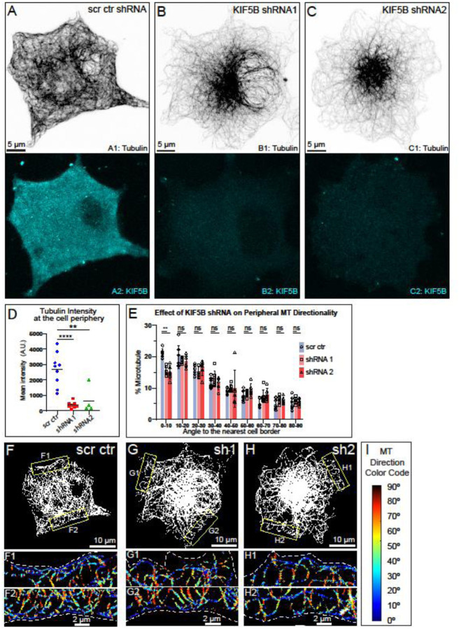
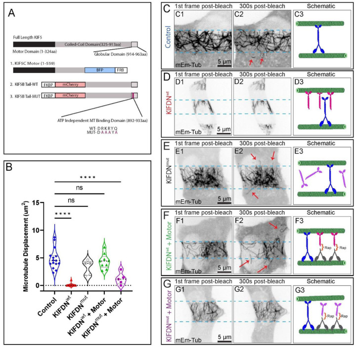
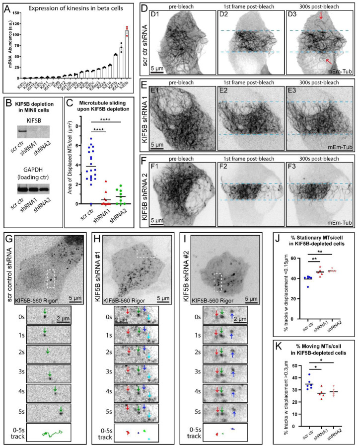
In pancreatic islet β cells, molecular motors use cytoskeletal polymers microtubules as tracks for intracellular transport of insulin secretory granules. The β-cell microtubule network has a complex architecture and is non-directional, which provides insulin granules at the cell periphery for rapid secretion response, yet to avoid over-secretion and subsequent hypoglycemia. We have previously characterized a peripheral sub-membrane microtubule array, which is critical for the withdrawal of excessive insulin granules from the secretion sites. Microtubules in β cells originate at the Golgi in the cell interior, and how the peripheral array is formed is unknown. Using real-time imaging and photo-kinetics approaches in clonal mouse pancreatic β cells MIN6, we now demonstrate that kinesin KIF5B, a motor protein with a capacity to transport microtubules as cargos, slides existing microtubules to the cell periphery and aligns them to each other along the plasma membrane. Moreover, like many physiological β-cell features, microtubule sliding is facilitated by a high glucose stimulus. These new data, together with our previous report that in high glucose sub-membrane MT array is destabilized to allow for robust secretion, indicate that MT sliding is another integral part of glucose-triggered microtubule remodeling, likely replacing destabilized peripheral microtubules to prevent their loss over time and β-cell malfunction.

 求助内容:
求助内容: 应助结果提醒方式:


