Joseph R Merrill, Alessandra Inguscio, Taemoon Chung, Breanna Demestichas, Libia A Garcia, Jill Habel, David Y Lewis, Tobias Janowitz, Scott K Lyons
{"title":"对癌症转移和免疫疗法反应进行灵敏、非免疫原性的体内成像。","authors":"Joseph R Merrill, Alessandra Inguscio, Taemoon Chung, Breanna Demestichas, Libia A Garcia, Jill Habel, David Y Lewis, Tobias Janowitz, Scott K Lyons","doi":"10.15698/cst2023.08.288","DOIUrl":null,"url":null,"abstract":"<p><p>Non-invasive imaging of tumors expressing reporter transgenes is a popular preclinical method for studying tumor development and response to therapy <i>in vivo</i> due to its ability to distinguish signal from tumors over background noise. However, the utilized transgenes, such as firefly luciferase, are immunogenic and, therefore, impact results when expressed in immune-competent hosts. This represents an important limitation, given that cancer immunology and immunotherapy are currently among the most impactful areas of research and therapeutic development. Here we present a non-immunogenic preclinical tumor imaging approach. Based on the expression of murine sodium iodide symporter (mNIS), it facilitates sensitive, non-invasive detection of syngeneic tumor cells in immune-competent tumor models without additional immunogenicity arising from exogenous transgenic protein or selection marker expression. NIS-expressing tumor cells internalize the gamma-emitting [<sup>99m</sup>Tc]pertechnetate ion and so can be detected by SPECT (single photon emission computed tomography). Using a mouse model of pancreatic ductal adenocarcinoma hepatic metastases in immune-competent C57BL/6 mice, we demonstrate that the technique enables the detection of very early metastatic lesions and longitudinal assessment of immunotherapy responses using precise and quantifiable whole-body SPECT/CT imaging.</p>","PeriodicalId":36371,"journal":{"name":"Cell Stress","volume":"7 8","pages":"59-68"},"PeriodicalIF":3.0000,"publicationDate":"2023-08-14","publicationTypes":"Journal Article","fieldsOfStudy":null,"isOpenAccess":false,"openAccessPdf":"https://www.ncbi.nlm.nih.gov/pmc/articles/PMC10468692/pdf/","citationCount":"0","resultStr":"{\"title\":\"Sensitive, non-immunogenic <i>in vivo</i> imaging of cancer metastases and immunotherapy response.\",\"authors\":\"Joseph R Merrill, Alessandra Inguscio, Taemoon Chung, Breanna Demestichas, Libia A Garcia, Jill Habel, David Y Lewis, Tobias Janowitz, Scott K Lyons\",\"doi\":\"10.15698/cst2023.08.288\",\"DOIUrl\":null,\"url\":null,\"abstract\":\"<p><p>Non-invasive imaging of tumors expressing reporter transgenes is a popular preclinical method for studying tumor development and response to therapy <i>in vivo</i> due to its ability to distinguish signal from tumors over background noise. However, the utilized transgenes, such as firefly luciferase, are immunogenic and, therefore, impact results when expressed in immune-competent hosts. This represents an important limitation, given that cancer immunology and immunotherapy are currently among the most impactful areas of research and therapeutic development. Here we present a non-immunogenic preclinical tumor imaging approach. Based on the expression of murine sodium iodide symporter (mNIS), it facilitates sensitive, non-invasive detection of syngeneic tumor cells in immune-competent tumor models without additional immunogenicity arising from exogenous transgenic protein or selection marker expression. NIS-expressing tumor cells internalize the gamma-emitting [<sup>99m</sup>Tc]pertechnetate ion and so can be detected by SPECT (single photon emission computed tomography). Using a mouse model of pancreatic ductal adenocarcinoma hepatic metastases in immune-competent C57BL/6 mice, we demonstrate that the technique enables the detection of very early metastatic lesions and longitudinal assessment of immunotherapy responses using precise and quantifiable whole-body SPECT/CT imaging.</p>\",\"PeriodicalId\":36371,\"journal\":{\"name\":\"Cell Stress\",\"volume\":\"7 8\",\"pages\":\"59-68\"},\"PeriodicalIF\":3.0000,\"publicationDate\":\"2023-08-14\",\"publicationTypes\":\"Journal Article\",\"fieldsOfStudy\":null,\"isOpenAccess\":false,\"openAccessPdf\":\"https://www.ncbi.nlm.nih.gov/pmc/articles/PMC10468692/pdf/\",\"citationCount\":\"0\",\"resultStr\":null,\"platform\":\"Semanticscholar\",\"paperid\":null,\"PeriodicalName\":\"Cell Stress\",\"FirstCategoryId\":\"1085\",\"ListUrlMain\":\"https://doi.org/10.15698/cst2023.08.288\",\"RegionNum\":0,\"RegionCategory\":null,\"ArticlePicture\":[],\"TitleCN\":null,\"AbstractTextCN\":null,\"PMCID\":null,\"EPubDate\":\"\",\"PubModel\":\"\",\"JCR\":\"Q2\",\"JCRName\":\"CELL BIOLOGY\",\"Score\":null,\"Total\":0}","platform":"Semanticscholar","paperid":null,"PeriodicalName":"Cell Stress","FirstCategoryId":"1085","ListUrlMain":"https://doi.org/10.15698/cst2023.08.288","RegionNum":0,"RegionCategory":null,"ArticlePicture":[],"TitleCN":null,"AbstractTextCN":null,"PMCID":null,"EPubDate":"","PubModel":"","JCR":"Q2","JCRName":"CELL BIOLOGY","Score":null,"Total":0}
Sensitive, non-immunogenic in vivo imaging of cancer metastases and immunotherapy response.
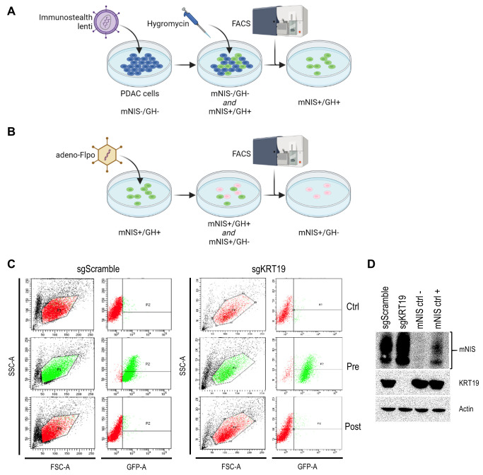
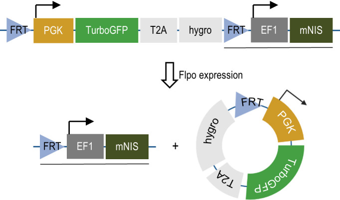
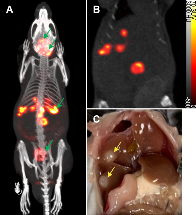
Non-invasive imaging of tumors expressing reporter transgenes is a popular preclinical method for studying tumor development and response to therapy in vivo due to its ability to distinguish signal from tumors over background noise. However, the utilized transgenes, such as firefly luciferase, are immunogenic and, therefore, impact results when expressed in immune-competent hosts. This represents an important limitation, given that cancer immunology and immunotherapy are currently among the most impactful areas of research and therapeutic development. Here we present a non-immunogenic preclinical tumor imaging approach. Based on the expression of murine sodium iodide symporter (mNIS), it facilitates sensitive, non-invasive detection of syngeneic tumor cells in immune-competent tumor models without additional immunogenicity arising from exogenous transgenic protein or selection marker expression. NIS-expressing tumor cells internalize the gamma-emitting [99mTc]pertechnetate ion and so can be detected by SPECT (single photon emission computed tomography). Using a mouse model of pancreatic ductal adenocarcinoma hepatic metastases in immune-competent C57BL/6 mice, we demonstrate that the technique enables the detection of very early metastatic lesions and longitudinal assessment of immunotherapy responses using precise and quantifiable whole-body SPECT/CT imaging.
Cell StressBiochemistry, Genetics and Molecular Biology-Biochemistry, Genetics and Molecular Biology (miscellaneous)
CiteScore
13.50
自引率
0.00%
发文量
21
审稿时长
15 weeks
期刊介绍:
Cell Stress is an open-access, peer-reviewed journal that is dedicated to publishing highly relevant research in the field of cellular pathology. The journal focuses on advancing our understanding of the molecular, mechanistic, phenotypic, and other critical aspects that underpin cellular dysfunction and disease. It specifically aims to foster cell biology research that is applicable to a range of significant human diseases, including neurodegenerative disorders, myopathies, mitochondriopathies, infectious diseases, cancer, and pathological aging.
The scope of Cell Stress is broad, welcoming submissions that represent a spectrum of research from fundamental to translational and clinical studies. The journal is a valuable resource for scientists, educators, and policymakers worldwide, as well as for any individual with an interest in cellular pathology. It serves as a platform for the dissemination of research findings that are instrumental in the investigation, classification, diagnosis, and therapeutic management of major diseases. By being open-access, Cell Stress ensures that its content is freely available to a global audience, thereby promoting international scientific collaboration and accelerating the exchange of knowledge within the research community.
 求助内容:
求助内容: 应助结果提醒方式:


