Lei Zhang, Ting Lan, Junyao Chen, Zidong Wei, Houyin Shi, Guoyou Wang
{"title":"MRI上距腓前韧带-距腓后韧带角度的增加可能有助于评估慢性踝关节不稳定。","authors":"Lei Zhang, Ting Lan, Junyao Chen, Zidong Wei, Houyin Shi, Guoyou Wang","doi":"10.1007/s00276-023-03196-7","DOIUrl":null,"url":null,"abstract":"<p><strong>Purpose: </strong>This study intended to compare the difference between the anterior talofibular ligament (ATFL) and posterior talofibular ligament (PTFL) angle with chronic ankle instability (CAI) patients and healthy volunteers, and to confirm whether using the ATFL-PTFL angle could be a reliable assessment method for CAI, so as to improve the accuracy and specificity of clinical diagnosis.</p><p><strong>Methods: </strong>This retrospective study included 240 participants: 120 CAI patients and 120 healthy volunteers between 2015 and 2021. The ATFL-PTFL angle of the ankle region was gaged in the cross-sectional supine position on MRI between two groups. After participants undergoing a comprehensive MRI scanning, ATFL-PTFL angles were regarded as the main indicator of patients with the injured ATFLs and healthy volunteers to compare, and were measured by an experienced musculoskeletal radiologist. Moreover, other qualitative and quantitative indicators referring to anatomical and morphological characteristics of the AFTL were included in this study with MRI, such as the length, width, thickness, shape, continuity, and signal intensity of the ATFL, which can be used as secondary indicators.</p><p><strong>Results: </strong>In the CAI group, the ATFL-PTFL angle was 90.8° ± 5.7°, which was significantly different from the non-CAI group where the ATFL-PTFL angle for 80.0° ± 3.7° (p < 0.001). As for the ATFL-MRI characteristics, the length (p = 0.003), width (p < 0.001), and thickness (p < 0.001) in the CAI group were also significantly different from the non-CAI group. Over 90% of the cases, patients of the CAI group had injured ATFL with an irregular shape, non-continuous, and high or mixed signal intensity.</p><p><strong>Conclusion: </strong>Compared with healthy people, the ATFL-PTFL angle of most CAI patients is larger, which can be used as a secondary index to diagnose CAI. However, the MRI characteristic changes of ATFL may not relate to the increased ATFL-PTFL angle.</p>","PeriodicalId":49296,"journal":{"name":"Surgical and Radiologic Anatomy","volume":" ","pages":"1205-1211"},"PeriodicalIF":1.2000,"publicationDate":"2023-10-01","publicationTypes":"Journal Article","fieldsOfStudy":null,"isOpenAccess":false,"openAccessPdf":"https://www.ncbi.nlm.nih.gov/pmc/articles/PMC10533641/pdf/","citationCount":"0","resultStr":"{\"title\":\"The increased anterior talofibular ligament-posterior talofibular ligament angle on MRI may help evaluate chronic ankle instability.\",\"authors\":\"Lei Zhang, Ting Lan, Junyao Chen, Zidong Wei, Houyin Shi, Guoyou Wang\",\"doi\":\"10.1007/s00276-023-03196-7\",\"DOIUrl\":null,\"url\":null,\"abstract\":\"<p><strong>Purpose: </strong>This study intended to compare the difference between the anterior talofibular ligament (ATFL) and posterior talofibular ligament (PTFL) angle with chronic ankle instability (CAI) patients and healthy volunteers, and to confirm whether using the ATFL-PTFL angle could be a reliable assessment method for CAI, so as to improve the accuracy and specificity of clinical diagnosis.</p><p><strong>Methods: </strong>This retrospective study included 240 participants: 120 CAI patients and 120 healthy volunteers between 2015 and 2021. The ATFL-PTFL angle of the ankle region was gaged in the cross-sectional supine position on MRI between two groups. After participants undergoing a comprehensive MRI scanning, ATFL-PTFL angles were regarded as the main indicator of patients with the injured ATFLs and healthy volunteers to compare, and were measured by an experienced musculoskeletal radiologist. Moreover, other qualitative and quantitative indicators referring to anatomical and morphological characteristics of the AFTL were included in this study with MRI, such as the length, width, thickness, shape, continuity, and signal intensity of the ATFL, which can be used as secondary indicators.</p><p><strong>Results: </strong>In the CAI group, the ATFL-PTFL angle was 90.8° ± 5.7°, which was significantly different from the non-CAI group where the ATFL-PTFL angle for 80.0° ± 3.7° (p < 0.001). As for the ATFL-MRI characteristics, the length (p = 0.003), width (p < 0.001), and thickness (p < 0.001) in the CAI group were also significantly different from the non-CAI group. Over 90% of the cases, patients of the CAI group had injured ATFL with an irregular shape, non-continuous, and high or mixed signal intensity.</p><p><strong>Conclusion: </strong>Compared with healthy people, the ATFL-PTFL angle of most CAI patients is larger, which can be used as a secondary index to diagnose CAI. However, the MRI characteristic changes of ATFL may not relate to the increased ATFL-PTFL angle.</p>\",\"PeriodicalId\":49296,\"journal\":{\"name\":\"Surgical and Radiologic Anatomy\",\"volume\":\" \",\"pages\":\"1205-1211\"},\"PeriodicalIF\":1.2000,\"publicationDate\":\"2023-10-01\",\"publicationTypes\":\"Journal Article\",\"fieldsOfStudy\":null,\"isOpenAccess\":false,\"openAccessPdf\":\"https://www.ncbi.nlm.nih.gov/pmc/articles/PMC10533641/pdf/\",\"citationCount\":\"0\",\"resultStr\":null,\"platform\":\"Semanticscholar\",\"paperid\":null,\"PeriodicalName\":\"Surgical and Radiologic Anatomy\",\"FirstCategoryId\":\"3\",\"ListUrlMain\":\"https://doi.org/10.1007/s00276-023-03196-7\",\"RegionNum\":4,\"RegionCategory\":\"医学\",\"ArticlePicture\":[],\"TitleCN\":null,\"AbstractTextCN\":null,\"PMCID\":null,\"EPubDate\":\"2023/7/10 0:00:00\",\"PubModel\":\"Epub\",\"JCR\":\"Q3\",\"JCRName\":\"ANATOMY & MORPHOLOGY\",\"Score\":null,\"Total\":0}","platform":"Semanticscholar","paperid":null,"PeriodicalName":"Surgical and Radiologic Anatomy","FirstCategoryId":"3","ListUrlMain":"https://doi.org/10.1007/s00276-023-03196-7","RegionNum":4,"RegionCategory":"医学","ArticlePicture":[],"TitleCN":null,"AbstractTextCN":null,"PMCID":null,"EPubDate":"2023/7/10 0:00:00","PubModel":"Epub","JCR":"Q3","JCRName":"ANATOMY & MORPHOLOGY","Score":null,"Total":0}
The increased anterior talofibular ligament-posterior talofibular ligament angle on MRI may help evaluate chronic ankle instability.
Purpose: This study intended to compare the difference between the anterior talofibular ligament (ATFL) and posterior talofibular ligament (PTFL) angle with chronic ankle instability (CAI) patients and healthy volunteers, and to confirm whether using the ATFL-PTFL angle could be a reliable assessment method for CAI, so as to improve the accuracy and specificity of clinical diagnosis.
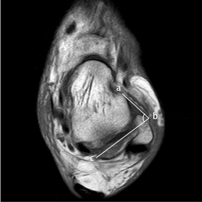
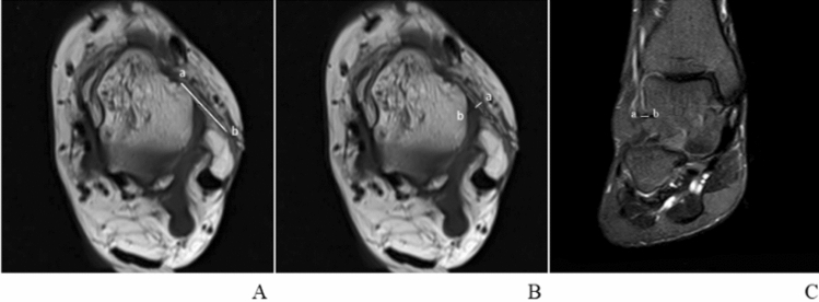
Methods: This retrospective study included 240 participants: 120 CAI patients and 120 healthy volunteers between 2015 and 2021. The ATFL-PTFL angle of the ankle region was gaged in the cross-sectional supine position on MRI between two groups. After participants undergoing a comprehensive MRI scanning, ATFL-PTFL angles were regarded as the main indicator of patients with the injured ATFLs and healthy volunteers to compare, and were measured by an experienced musculoskeletal radiologist. Moreover, other qualitative and quantitative indicators referring to anatomical and morphological characteristics of the AFTL were included in this study with MRI, such as the length, width, thickness, shape, continuity, and signal intensity of the ATFL, which can be used as secondary indicators.
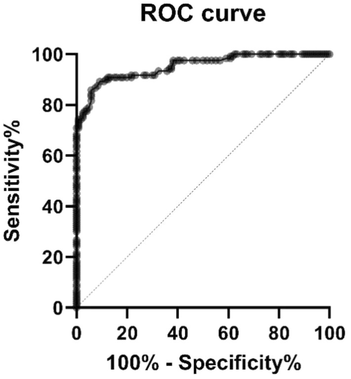
Results: In the CAI group, the ATFL-PTFL angle was 90.8° ± 5.7°, which was significantly different from the non-CAI group where the ATFL-PTFL angle for 80.0° ± 3.7° (p < 0.001). As for the ATFL-MRI characteristics, the length (p = 0.003), width (p < 0.001), and thickness (p < 0.001) in the CAI group were also significantly different from the non-CAI group. Over 90% of the cases, patients of the CAI group had injured ATFL with an irregular shape, non-continuous, and high or mixed signal intensity.
Conclusion: Compared with healthy people, the ATFL-PTFL angle of most CAI patients is larger, which can be used as a secondary index to diagnose CAI. However, the MRI characteristic changes of ATFL may not relate to the increased ATFL-PTFL angle.
期刊介绍:
Anatomy is a morphological science which cannot fail to interest the clinician. The practical application of anatomical research to clinical problems necessitates special adaptation and selectivity in choosing from numerous international works. Although there is a tendency to believe that meaningful advances in anatomy are unlikely, constant revision is necessary. Surgical and Radiologic Anatomy, the first international journal of Clinical anatomy has been created in this spirit.
Its goal is to serve clinicians, regardless of speciality-physicians, surgeons, radiologists or other specialists-as an indispensable aid with which they can improve their knowledge of anatomy. Each issue includes: Original papers, review articles, articles on the anatomical bases of medical, surgical and radiological techniques, articles of normal radiologic anatomy, brief reviews of anatomical publications of clinical interest.
Particular attention is given to high quality illustrations, which are indispensable for a better understanding of anatomical problems.
Surgical and Radiologic Anatomy is a journal written by anatomists for clinicians with a special interest in anatomy.

 求助内容:
求助内容: 应助结果提醒方式:


