{"title":"三叉神经恶性黑色素神经鞘瘤1例报告及文献复习。","authors":"Anurag Chandrakant Dandekar, Nirav A Mehta","doi":"10.1055/s-0043-1768578","DOIUrl":null,"url":null,"abstract":"<p><p>Intracranial melanotic schwannoma is quite rare, and involvement of the trigeminal nerve is even rarer. Early diagnosis and surgical excision are the mainstays of management. These tumors have a high tendency to recur and there is high possibility of metastasis. Adjuvant radiotherapy should be considered since the prognosis is uncertain. A 23-year-old man started developing numbness over the left side of the forehead 9 months ago that progressed to involve the ipsilateral cheek. The patient started having diplopia on looking to the left side 8 months ago. His relatives noticed a change in his voice 1 month ago and he developed weakness in the right upper and lower limbs, which was gradually progressive. The patient had slight difficulty swallowing. After examination, we found involvement of multiple cranial nerves with pyramidal signs. Magnetic resonance imaging (MRI) was suggestive of an extra-axial lesion in the left cerebellopontine angle extending into the middle cranial fossa, which was having high T1 and T2 signal loss with contrast enhancement. We achieved near-total excision of the tumor via a subtemporal extradural approach. Trigeminal melanotic schwannoma is a rare occurrence constituting melanin-producing cells and Schwann cells. Rapid progression of symptoms and signs should prompt the suspicion of the possible malignant nature of the pathology. Extradural skull base approaches reduce the risk of postoperative deficits. Differentiating melanotic schwannoma from malignant melanoma is of utmost importance in planning of management.</p>","PeriodicalId":8521,"journal":{"name":"Asian Journal of Neurosurgery","volume":"18 2","pages":"352-356"},"PeriodicalIF":0.0000,"publicationDate":"2023-06-01","publicationTypes":"Journal Article","fieldsOfStudy":null,"isOpenAccess":false,"openAccessPdf":"https://ftp.ncbi.nlm.nih.gov/pub/pmc/oa_pdf/80/8b/10-1055-s-0043-1768578.PMC10313432.pdf","citationCount":"0","resultStr":"{\"title\":\"A Case of Malignant Melanotic Schwannoma of the Trigeminal Nerve: A Case Report and Review of Literature.\",\"authors\":\"Anurag Chandrakant Dandekar, Nirav A Mehta\",\"doi\":\"10.1055/s-0043-1768578\",\"DOIUrl\":null,\"url\":null,\"abstract\":\"<p><p>Intracranial melanotic schwannoma is quite rare, and involvement of the trigeminal nerve is even rarer. Early diagnosis and surgical excision are the mainstays of management. These tumors have a high tendency to recur and there is high possibility of metastasis. Adjuvant radiotherapy should be considered since the prognosis is uncertain. A 23-year-old man started developing numbness over the left side of the forehead 9 months ago that progressed to involve the ipsilateral cheek. The patient started having diplopia on looking to the left side 8 months ago. His relatives noticed a change in his voice 1 month ago and he developed weakness in the right upper and lower limbs, which was gradually progressive. The patient had slight difficulty swallowing. After examination, we found involvement of multiple cranial nerves with pyramidal signs. Magnetic resonance imaging (MRI) was suggestive of an extra-axial lesion in the left cerebellopontine angle extending into the middle cranial fossa, which was having high T1 and T2 signal loss with contrast enhancement. We achieved near-total excision of the tumor via a subtemporal extradural approach. Trigeminal melanotic schwannoma is a rare occurrence constituting melanin-producing cells and Schwann cells. Rapid progression of symptoms and signs should prompt the suspicion of the possible malignant nature of the pathology. Extradural skull base approaches reduce the risk of postoperative deficits. Differentiating melanotic schwannoma from malignant melanoma is of utmost importance in planning of management.</p>\",\"PeriodicalId\":8521,\"journal\":{\"name\":\"Asian Journal of Neurosurgery\",\"volume\":\"18 2\",\"pages\":\"352-356\"},\"PeriodicalIF\":0.0000,\"publicationDate\":\"2023-06-01\",\"publicationTypes\":\"Journal Article\",\"fieldsOfStudy\":null,\"isOpenAccess\":false,\"openAccessPdf\":\"https://ftp.ncbi.nlm.nih.gov/pub/pmc/oa_pdf/80/8b/10-1055-s-0043-1768578.PMC10313432.pdf\",\"citationCount\":\"0\",\"resultStr\":null,\"platform\":\"Semanticscholar\",\"paperid\":null,\"PeriodicalName\":\"Asian Journal of Neurosurgery\",\"FirstCategoryId\":\"1085\",\"ListUrlMain\":\"https://doi.org/10.1055/s-0043-1768578\",\"RegionNum\":0,\"RegionCategory\":null,\"ArticlePicture\":[],\"TitleCN\":null,\"AbstractTextCN\":null,\"PMCID\":null,\"EPubDate\":\"\",\"PubModel\":\"\",\"JCR\":\"\",\"JCRName\":\"\",\"Score\":null,\"Total\":0}","platform":"Semanticscholar","paperid":null,"PeriodicalName":"Asian Journal of Neurosurgery","FirstCategoryId":"1085","ListUrlMain":"https://doi.org/10.1055/s-0043-1768578","RegionNum":0,"RegionCategory":null,"ArticlePicture":[],"TitleCN":null,"AbstractTextCN":null,"PMCID":null,"EPubDate":"","PubModel":"","JCR":"","JCRName":"","Score":null,"Total":0}
A Case of Malignant Melanotic Schwannoma of the Trigeminal Nerve: A Case Report and Review of Literature.
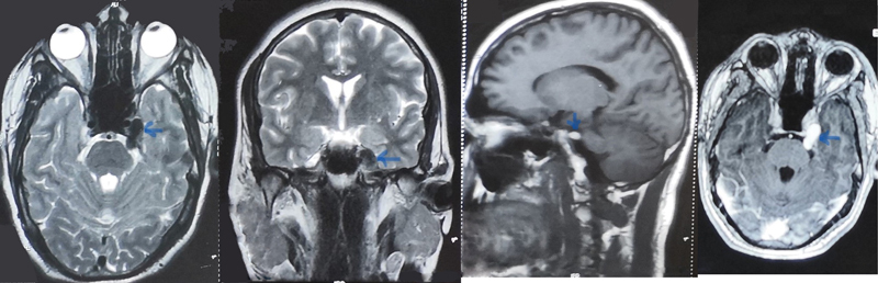
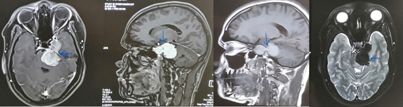
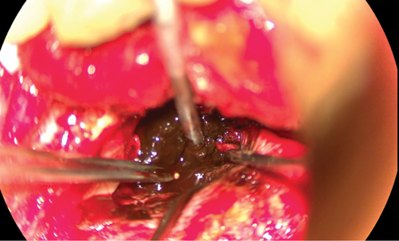
Intracranial melanotic schwannoma is quite rare, and involvement of the trigeminal nerve is even rarer. Early diagnosis and surgical excision are the mainstays of management. These tumors have a high tendency to recur and there is high possibility of metastasis. Adjuvant radiotherapy should be considered since the prognosis is uncertain. A 23-year-old man started developing numbness over the left side of the forehead 9 months ago that progressed to involve the ipsilateral cheek. The patient started having diplopia on looking to the left side 8 months ago. His relatives noticed a change in his voice 1 month ago and he developed weakness in the right upper and lower limbs, which was gradually progressive. The patient had slight difficulty swallowing. After examination, we found involvement of multiple cranial nerves with pyramidal signs. Magnetic resonance imaging (MRI) was suggestive of an extra-axial lesion in the left cerebellopontine angle extending into the middle cranial fossa, which was having high T1 and T2 signal loss with contrast enhancement. We achieved near-total excision of the tumor via a subtemporal extradural approach. Trigeminal melanotic schwannoma is a rare occurrence constituting melanin-producing cells and Schwann cells. Rapid progression of symptoms and signs should prompt the suspicion of the possible malignant nature of the pathology. Extradural skull base approaches reduce the risk of postoperative deficits. Differentiating melanotic schwannoma from malignant melanoma is of utmost importance in planning of management.

 求助内容:
求助内容: 应助结果提醒方式:


