Kyu-Ho Yi, Hyung-Jin Lee, Ji-Hyun Lee, Min Ho An, Kangwoo Lee, Hyewon Hu, Min-Seung Kim, Hosung Choi, Hee-Jin Kim
{"title":"颈阔带的声解剖:颈阔带是什么原因引起的?","authors":"Kyu-Ho Yi, Hyung-Jin Lee, Ji-Hyun Lee, Min Ho An, Kangwoo Lee, Hyewon Hu, Min-Seung Kim, Hosung Choi, Hee-Jin Kim","doi":"10.1007/s00276-023-03236-2","DOIUrl":null,"url":null,"abstract":"<p><strong>Background: </strong>The platysmal band is created by the platysma muscle, a thin superficial muscle that covers the entire neck and the lower part of the face. The platysmal band appears at the anterior and posterior borders of the muscle. To date, no definite pathophysiology has been established. Here, we observed a lack of knowledge of the anatomy of the platysma muscle using ultrasonography in this study.</p><p><strong>Methods: </strong>We conducted a descriptive, prospective study observing the platysmal band in resting and contraction states to reveal muscle changes. Twenty-four participants (aged 23-57 years) with anterior and posterior neck bands underwent ultrasonography in resting and contracted states. Ten cadavers were studied aged 67-85 years to measure the thickness of the platysma muscle at 12 points: horizontally (medial, middle, lateral) and vertically (inferior mandibular margin, hyoid bone, cricoid cartilage, superior margin of clavicle).</p><p><strong>Results: </strong>The anterior and posterior borders of the platysma muscle were thicker than the middle of the platysma muscle when in a contracted state, and the muscle also had a convex shape when contracted. The thickness of the platysma muscle was not significantly different over 12 points in the resting state. During contraction, the platysma muscles contracted in the medial and lateral margins of the muscle, which was more significant in the posterior bands.</p><p><strong>Conclusion: </strong>The anterior and posterior platysmal bands are related to muscle thickness during contraction. These observations support the change in platysmal band treatment only at the anterior and posterior border of the muscle.</p>","PeriodicalId":49296,"journal":{"name":"Surgical and Radiologic Anatomy","volume":" ","pages":"1399-1404"},"PeriodicalIF":1.2000,"publicationDate":"2023-11-01","publicationTypes":"Journal Article","fieldsOfStudy":null,"isOpenAccess":false,"openAccessPdf":"","citationCount":"0","resultStr":"{\"title\":\"Sonoanatomy of the platysmal bands: What causes the platysmal band?\",\"authors\":\"Kyu-Ho Yi, Hyung-Jin Lee, Ji-Hyun Lee, Min Ho An, Kangwoo Lee, Hyewon Hu, Min-Seung Kim, Hosung Choi, Hee-Jin Kim\",\"doi\":\"10.1007/s00276-023-03236-2\",\"DOIUrl\":null,\"url\":null,\"abstract\":\"<p><strong>Background: </strong>The platysmal band is created by the platysma muscle, a thin superficial muscle that covers the entire neck and the lower part of the face. The platysmal band appears at the anterior and posterior borders of the muscle. To date, no definite pathophysiology has been established. Here, we observed a lack of knowledge of the anatomy of the platysma muscle using ultrasonography in this study.</p><p><strong>Methods: </strong>We conducted a descriptive, prospective study observing the platysmal band in resting and contraction states to reveal muscle changes. Twenty-four participants (aged 23-57 years) with anterior and posterior neck bands underwent ultrasonography in resting and contracted states. Ten cadavers were studied aged 67-85 years to measure the thickness of the platysma muscle at 12 points: horizontally (medial, middle, lateral) and vertically (inferior mandibular margin, hyoid bone, cricoid cartilage, superior margin of clavicle).</p><p><strong>Results: </strong>The anterior and posterior borders of the platysma muscle were thicker than the middle of the platysma muscle when in a contracted state, and the muscle also had a convex shape when contracted. The thickness of the platysma muscle was not significantly different over 12 points in the resting state. During contraction, the platysma muscles contracted in the medial and lateral margins of the muscle, which was more significant in the posterior bands.</p><p><strong>Conclusion: </strong>The anterior and posterior platysmal bands are related to muscle thickness during contraction. These observations support the change in platysmal band treatment only at the anterior and posterior border of the muscle.</p>\",\"PeriodicalId\":49296,\"journal\":{\"name\":\"Surgical and Radiologic Anatomy\",\"volume\":\" \",\"pages\":\"1399-1404\"},\"PeriodicalIF\":1.2000,\"publicationDate\":\"2023-11-01\",\"publicationTypes\":\"Journal Article\",\"fieldsOfStudy\":null,\"isOpenAccess\":false,\"openAccessPdf\":\"\",\"citationCount\":\"0\",\"resultStr\":null,\"platform\":\"Semanticscholar\",\"paperid\":null,\"PeriodicalName\":\"Surgical and Radiologic Anatomy\",\"FirstCategoryId\":\"3\",\"ListUrlMain\":\"https://doi.org/10.1007/s00276-023-03236-2\",\"RegionNum\":4,\"RegionCategory\":\"医学\",\"ArticlePicture\":[],\"TitleCN\":null,\"AbstractTextCN\":null,\"PMCID\":null,\"EPubDate\":\"2023/8/29 0:00:00\",\"PubModel\":\"Epub\",\"JCR\":\"Q3\",\"JCRName\":\"ANATOMY & MORPHOLOGY\",\"Score\":null,\"Total\":0}","platform":"Semanticscholar","paperid":null,"PeriodicalName":"Surgical and Radiologic Anatomy","FirstCategoryId":"3","ListUrlMain":"https://doi.org/10.1007/s00276-023-03236-2","RegionNum":4,"RegionCategory":"医学","ArticlePicture":[],"TitleCN":null,"AbstractTextCN":null,"PMCID":null,"EPubDate":"2023/8/29 0:00:00","PubModel":"Epub","JCR":"Q3","JCRName":"ANATOMY & MORPHOLOGY","Score":null,"Total":0}
Sonoanatomy of the platysmal bands: What causes the platysmal band?
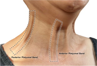
Background: The platysmal band is created by the platysma muscle, a thin superficial muscle that covers the entire neck and the lower part of the face. The platysmal band appears at the anterior and posterior borders of the muscle. To date, no definite pathophysiology has been established. Here, we observed a lack of knowledge of the anatomy of the platysma muscle using ultrasonography in this study.
Methods: We conducted a descriptive, prospective study observing the platysmal band in resting and contraction states to reveal muscle changes. Twenty-four participants (aged 23-57 years) with anterior and posterior neck bands underwent ultrasonography in resting and contracted states. Ten cadavers were studied aged 67-85 years to measure the thickness of the platysma muscle at 12 points: horizontally (medial, middle, lateral) and vertically (inferior mandibular margin, hyoid bone, cricoid cartilage, superior margin of clavicle).
Results: The anterior and posterior borders of the platysma muscle were thicker than the middle of the platysma muscle when in a contracted state, and the muscle also had a convex shape when contracted. The thickness of the platysma muscle was not significantly different over 12 points in the resting state. During contraction, the platysma muscles contracted in the medial and lateral margins of the muscle, which was more significant in the posterior bands.
Conclusion: The anterior and posterior platysmal bands are related to muscle thickness during contraction. These observations support the change in platysmal band treatment only at the anterior and posterior border of the muscle.
期刊介绍:
Anatomy is a morphological science which cannot fail to interest the clinician. The practical application of anatomical research to clinical problems necessitates special adaptation and selectivity in choosing from numerous international works. Although there is a tendency to believe that meaningful advances in anatomy are unlikely, constant revision is necessary. Surgical and Radiologic Anatomy, the first international journal of Clinical anatomy has been created in this spirit.
Its goal is to serve clinicians, regardless of speciality-physicians, surgeons, radiologists or other specialists-as an indispensable aid with which they can improve their knowledge of anatomy. Each issue includes: Original papers, review articles, articles on the anatomical bases of medical, surgical and radiological techniques, articles of normal radiologic anatomy, brief reviews of anatomical publications of clinical interest.
Particular attention is given to high quality illustrations, which are indispensable for a better understanding of anatomical problems.
Surgical and Radiologic Anatomy is a journal written by anatomists for clinicians with a special interest in anatomy.

 求助内容:
求助内容: 应助结果提醒方式:


