{"title":"胰背动脉和胰内窦解剖的MDCT评估。","authors":"Shaurya Sharma, Binit Sureka, Vaibhav Varshney, Subhash Soni, Taruna Yadav, Pawan Kumar Garg, Pushpinder Singh Khera","doi":"10.1007/s00276-023-03235-3","DOIUrl":null,"url":null,"abstract":"<p><strong>Objective: </strong>The purpose of the study was to analyze the anatomy and variations in the origin of the dorsal pancreatic artery, greater pancreatic artery and to study the various types of arterial arcades supplying the pancreas on multidetector CT (MDCT).</p><p><strong>Methods: </strong>A retrospective analysis of 747 MDCT scans was performed in patients who underwent triple phase or dual phase CT abdomen between December 2020 and October 2022. Variations in origin of Dorsal pancreatic artery (DPA), greater pancreatic artery (GPA), uncinate process branch were studied. Intrapancreatic arcade anatomy was classified according to Roman Ramos et al. into 4 types-small arcades (type I), small and large arcades (type II), large arcades (type III) and straight branches (type IV).</p><p><strong>Results: </strong>The DPA was visualized in 65.3% (n = 488) of cases. The most common origin was from the splenic artery in 58.2% (n = 284) cases. The mean calibre of DPA was 2.05 mm (1.0-4.8 mm). The uncinate branch was seen in 21.7% (n = 106) with an average diameter of 1.3 mm. The greater pancreatic artery was seen in 57.3% (n = 428) predominantly seen arising from the splenic artery. The most common arcade anatomy was of Type II in 52.1% (n = 63) cases.</p><p><strong>Conclusion: </strong>Pancreatic arterial variations are not very uncommon in daily practice. Knowledge of these variations before pancreatic surgery and endovascular intervention procedure is important for surgeons and interventional radiologist.</p>","PeriodicalId":49296,"journal":{"name":"Surgical and Radiologic Anatomy","volume":" ","pages":"1471-1476"},"PeriodicalIF":1.2000,"publicationDate":"2023-11-01","publicationTypes":"Journal Article","fieldsOfStudy":null,"isOpenAccess":false,"openAccessPdf":"","citationCount":"0","resultStr":"{\"title\":\"MDCT evaluation of Dorsal Pancreatic Artery and Intrapancreatic arcade anatomy.\",\"authors\":\"Shaurya Sharma, Binit Sureka, Vaibhav Varshney, Subhash Soni, Taruna Yadav, Pawan Kumar Garg, Pushpinder Singh Khera\",\"doi\":\"10.1007/s00276-023-03235-3\",\"DOIUrl\":null,\"url\":null,\"abstract\":\"<p><strong>Objective: </strong>The purpose of the study was to analyze the anatomy and variations in the origin of the dorsal pancreatic artery, greater pancreatic artery and to study the various types of arterial arcades supplying the pancreas on multidetector CT (MDCT).</p><p><strong>Methods: </strong>A retrospective analysis of 747 MDCT scans was performed in patients who underwent triple phase or dual phase CT abdomen between December 2020 and October 2022. Variations in origin of Dorsal pancreatic artery (DPA), greater pancreatic artery (GPA), uncinate process branch were studied. Intrapancreatic arcade anatomy was classified according to Roman Ramos et al. into 4 types-small arcades (type I), small and large arcades (type II), large arcades (type III) and straight branches (type IV).</p><p><strong>Results: </strong>The DPA was visualized in 65.3% (n = 488) of cases. The most common origin was from the splenic artery in 58.2% (n = 284) cases. The mean calibre of DPA was 2.05 mm (1.0-4.8 mm). The uncinate branch was seen in 21.7% (n = 106) with an average diameter of 1.3 mm. The greater pancreatic artery was seen in 57.3% (n = 428) predominantly seen arising from the splenic artery. The most common arcade anatomy was of Type II in 52.1% (n = 63) cases.</p><p><strong>Conclusion: </strong>Pancreatic arterial variations are not very uncommon in daily practice. Knowledge of these variations before pancreatic surgery and endovascular intervention procedure is important for surgeons and interventional radiologist.</p>\",\"PeriodicalId\":49296,\"journal\":{\"name\":\"Surgical and Radiologic Anatomy\",\"volume\":\" \",\"pages\":\"1471-1476\"},\"PeriodicalIF\":1.2000,\"publicationDate\":\"2023-11-01\",\"publicationTypes\":\"Journal Article\",\"fieldsOfStudy\":null,\"isOpenAccess\":false,\"openAccessPdf\":\"\",\"citationCount\":\"0\",\"resultStr\":null,\"platform\":\"Semanticscholar\",\"paperid\":null,\"PeriodicalName\":\"Surgical and Radiologic Anatomy\",\"FirstCategoryId\":\"3\",\"ListUrlMain\":\"https://doi.org/10.1007/s00276-023-03235-3\",\"RegionNum\":4,\"RegionCategory\":\"医学\",\"ArticlePicture\":[],\"TitleCN\":null,\"AbstractTextCN\":null,\"PMCID\":null,\"EPubDate\":\"2023/8/28 0:00:00\",\"PubModel\":\"Epub\",\"JCR\":\"Q3\",\"JCRName\":\"ANATOMY & MORPHOLOGY\",\"Score\":null,\"Total\":0}","platform":"Semanticscholar","paperid":null,"PeriodicalName":"Surgical and Radiologic Anatomy","FirstCategoryId":"3","ListUrlMain":"https://doi.org/10.1007/s00276-023-03235-3","RegionNum":4,"RegionCategory":"医学","ArticlePicture":[],"TitleCN":null,"AbstractTextCN":null,"PMCID":null,"EPubDate":"2023/8/28 0:00:00","PubModel":"Epub","JCR":"Q3","JCRName":"ANATOMY & MORPHOLOGY","Score":null,"Total":0}
MDCT evaluation of Dorsal Pancreatic Artery and Intrapancreatic arcade anatomy.
Objective: The purpose of the study was to analyze the anatomy and variations in the origin of the dorsal pancreatic artery, greater pancreatic artery and to study the various types of arterial arcades supplying the pancreas on multidetector CT (MDCT).
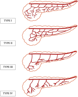
Methods: A retrospective analysis of 747 MDCT scans was performed in patients who underwent triple phase or dual phase CT abdomen between December 2020 and October 2022. Variations in origin of Dorsal pancreatic artery (DPA), greater pancreatic artery (GPA), uncinate process branch were studied. Intrapancreatic arcade anatomy was classified according to Roman Ramos et al. into 4 types-small arcades (type I), small and large arcades (type II), large arcades (type III) and straight branches (type IV).
Results: The DPA was visualized in 65.3% (n = 488) of cases. The most common origin was from the splenic artery in 58.2% (n = 284) cases. The mean calibre of DPA was 2.05 mm (1.0-4.8 mm). The uncinate branch was seen in 21.7% (n = 106) with an average diameter of 1.3 mm. The greater pancreatic artery was seen in 57.3% (n = 428) predominantly seen arising from the splenic artery. The most common arcade anatomy was of Type II in 52.1% (n = 63) cases.
Conclusion: Pancreatic arterial variations are not very uncommon in daily practice. Knowledge of these variations before pancreatic surgery and endovascular intervention procedure is important for surgeons and interventional radiologist.
期刊介绍:
Anatomy is a morphological science which cannot fail to interest the clinician. The practical application of anatomical research to clinical problems necessitates special adaptation and selectivity in choosing from numerous international works. Although there is a tendency to believe that meaningful advances in anatomy are unlikely, constant revision is necessary. Surgical and Radiologic Anatomy, the first international journal of Clinical anatomy has been created in this spirit.
Its goal is to serve clinicians, regardless of speciality-physicians, surgeons, radiologists or other specialists-as an indispensable aid with which they can improve their knowledge of anatomy. Each issue includes: Original papers, review articles, articles on the anatomical bases of medical, surgical and radiological techniques, articles of normal radiologic anatomy, brief reviews of anatomical publications of clinical interest.
Particular attention is given to high quality illustrations, which are indispensable for a better understanding of anatomical problems.
Surgical and Radiologic Anatomy is a journal written by anatomists for clinicians with a special interest in anatomy.

 求助内容:
求助内容: 应助结果提醒方式:


