{"title":"海绵窦硬脊膜动静脉瘘通过闭塞的岩上窦栓塞:一个例证性病例。","authors":"Natsuki Akaike, Hiroyuki Ikeda, Kensuke Takada, Minami Uezato, Masanori Kinosada, Yoshitaka Kurosaki, Masaki Chin","doi":"10.3171/CASE23143","DOIUrl":null,"url":null,"abstract":"<p><strong>Background: </strong>Transvenous embolization for cavernous sinus (CS) dural arteriovenous fistulas (CS-DAVFs) with limitations of the major access routes to the CS is challenging.</p><p><strong>Observations: </strong>A 74-year-old woman presented with left-sided conjunctival injection and exophthalmos. Cerebral angiography showed a left CS-DAVF draining into the left uncal vein and superior ophthalmic vein, with the fistulous point located in the posterosuperior compartment of the left CS. The left inferior petrosal sinus and internal jugular vein were occluded, and no drainage route from the left superior ophthalmic vein was seen. The anterior segment of the left superior petrosal sinus (SPS) was occluded, but the posterior segment was not. Microangiography from the posterior segment of the left SPS showed a beak-like orifice in the anterior segment of the left SPS toward the left CS. A micro-guidewire was guided through the beak-like orifice, and the microcatheter was advanced into the left CS. The left CS was packed and the DAVF was occluded.</p><p><strong>Lessons: </strong>Transvenous embolization through an occluded SPS may be an option in the endovascular treatment of CS-DAVFs. Penetration along the beak-like orifice of the occluded SPS visualized by venography at the blind end of the SPS may be useful in reaching the CS via the SPS.</p>","PeriodicalId":16554,"journal":{"name":"Journal of Neurosurgery: Case Lessons","volume":"5 25","pages":""},"PeriodicalIF":0.0000,"publicationDate":"2023-06-19","publicationTypes":"Journal Article","fieldsOfStudy":null,"isOpenAccess":false,"openAccessPdf":"https://ftp.ncbi.nlm.nih.gov/pub/pmc/oa_pdf/9e/e0/CASE23143.PMC10550532.pdf","citationCount":"0","resultStr":"{\"title\":\"Cavernous sinus dural arteriovenous fistula embolized through an occluded superior petrosal sinus: illustrative case.\",\"authors\":\"Natsuki Akaike, Hiroyuki Ikeda, Kensuke Takada, Minami Uezato, Masanori Kinosada, Yoshitaka Kurosaki, Masaki Chin\",\"doi\":\"10.3171/CASE23143\",\"DOIUrl\":null,\"url\":null,\"abstract\":\"<p><strong>Background: </strong>Transvenous embolization for cavernous sinus (CS) dural arteriovenous fistulas (CS-DAVFs) with limitations of the major access routes to the CS is challenging.</p><p><strong>Observations: </strong>A 74-year-old woman presented with left-sided conjunctival injection and exophthalmos. Cerebral angiography showed a left CS-DAVF draining into the left uncal vein and superior ophthalmic vein, with the fistulous point located in the posterosuperior compartment of the left CS. The left inferior petrosal sinus and internal jugular vein were occluded, and no drainage route from the left superior ophthalmic vein was seen. The anterior segment of the left superior petrosal sinus (SPS) was occluded, but the posterior segment was not. Microangiography from the posterior segment of the left SPS showed a beak-like orifice in the anterior segment of the left SPS toward the left CS. A micro-guidewire was guided through the beak-like orifice, and the microcatheter was advanced into the left CS. The left CS was packed and the DAVF was occluded.</p><p><strong>Lessons: </strong>Transvenous embolization through an occluded SPS may be an option in the endovascular treatment of CS-DAVFs. Penetration along the beak-like orifice of the occluded SPS visualized by venography at the blind end of the SPS may be useful in reaching the CS via the SPS.</p>\",\"PeriodicalId\":16554,\"journal\":{\"name\":\"Journal of Neurosurgery: Case Lessons\",\"volume\":\"5 25\",\"pages\":\"\"},\"PeriodicalIF\":0.0000,\"publicationDate\":\"2023-06-19\",\"publicationTypes\":\"Journal Article\",\"fieldsOfStudy\":null,\"isOpenAccess\":false,\"openAccessPdf\":\"https://ftp.ncbi.nlm.nih.gov/pub/pmc/oa_pdf/9e/e0/CASE23143.PMC10550532.pdf\",\"citationCount\":\"0\",\"resultStr\":null,\"platform\":\"Semanticscholar\",\"paperid\":null,\"PeriodicalName\":\"Journal of Neurosurgery: Case Lessons\",\"FirstCategoryId\":\"1085\",\"ListUrlMain\":\"https://doi.org/10.3171/CASE23143\",\"RegionNum\":0,\"RegionCategory\":null,\"ArticlePicture\":[],\"TitleCN\":null,\"AbstractTextCN\":null,\"PMCID\":null,\"EPubDate\":\"\",\"PubModel\":\"\",\"JCR\":\"\",\"JCRName\":\"\",\"Score\":null,\"Total\":0}","platform":"Semanticscholar","paperid":null,"PeriodicalName":"Journal of Neurosurgery: Case Lessons","FirstCategoryId":"1085","ListUrlMain":"https://doi.org/10.3171/CASE23143","RegionNum":0,"RegionCategory":null,"ArticlePicture":[],"TitleCN":null,"AbstractTextCN":null,"PMCID":null,"EPubDate":"","PubModel":"","JCR":"","JCRName":"","Score":null,"Total":0}
Cavernous sinus dural arteriovenous fistula embolized through an occluded superior petrosal sinus: illustrative case.
Background: Transvenous embolization for cavernous sinus (CS) dural arteriovenous fistulas (CS-DAVFs) with limitations of the major access routes to the CS is challenging.
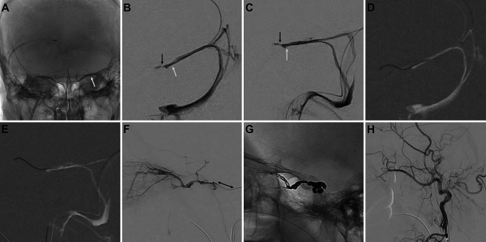
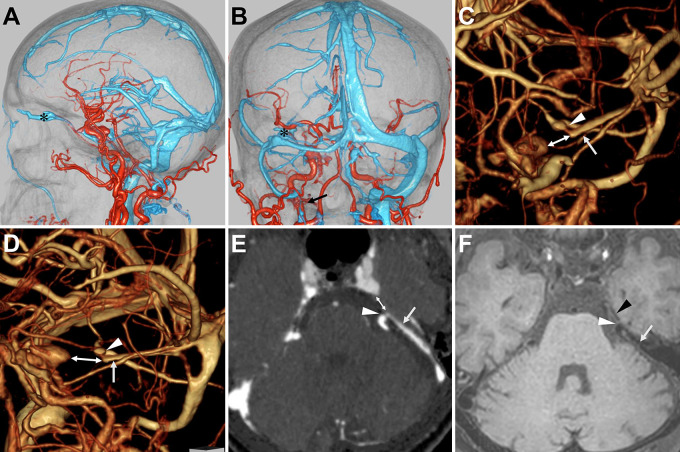
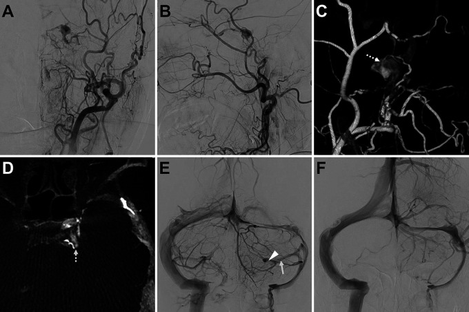
Observations: A 74-year-old woman presented with left-sided conjunctival injection and exophthalmos. Cerebral angiography showed a left CS-DAVF draining into the left uncal vein and superior ophthalmic vein, with the fistulous point located in the posterosuperior compartment of the left CS. The left inferior petrosal sinus and internal jugular vein were occluded, and no drainage route from the left superior ophthalmic vein was seen. The anterior segment of the left superior petrosal sinus (SPS) was occluded, but the posterior segment was not. Microangiography from the posterior segment of the left SPS showed a beak-like orifice in the anterior segment of the left SPS toward the left CS. A micro-guidewire was guided through the beak-like orifice, and the microcatheter was advanced into the left CS. The left CS was packed and the DAVF was occluded.
Lessons: Transvenous embolization through an occluded SPS may be an option in the endovascular treatment of CS-DAVFs. Penetration along the beak-like orifice of the occluded SPS visualized by venography at the blind end of the SPS may be useful in reaching the CS via the SPS.

 求助内容:
求助内容: 应助结果提醒方式:


