Hella Baumann, Melanie Schwingel, Marcello Sestu, Anna Burcza, Susanna Marg, Wolfgang Ziegler, Anna V. Taubenberger, Daniel J. Muller, Martin Bastmeyer, Clemens M. Franz
{"title":"血管蛋白对新生粘附的双相强化","authors":"Hella Baumann, Melanie Schwingel, Marcello Sestu, Anna Burcza, Susanna Marg, Wolfgang Ziegler, Anna V. Taubenberger, Daniel J. Muller, Martin Bastmeyer, Clemens M. Franz","doi":"10.1002/jmr.3012","DOIUrl":null,"url":null,"abstract":"<p>Vinculin is an integral component of integrin adhesions, where it functions as a molecular clutch coupling intracellular contraction to the extracellular matrix. Quantitating its contribution to the reinforcement of newly forming adhesions, however, requires ultrasensitive cell force assays covering short time and low force ranges. Here, we have combined atomic force microscopy-based single-cell force spectroscopy (SCFS) and optical tweezers force spectroscopy to investigate the role of vinculin in reinforcement of individual nascent adhesions during the first 5 min of cell contact with fibronectin or vitronectin. At minimal adhesion times (5-10 s), mouse embryonic fibroblast (MEF) wildtype (<i>wt</i>) and vinculin knock-out (<i>vin</i><sup>(−/−)</sup>) cells develop comparable adhesion forces on the scale of several individual integrin-ligand bonds, confirming that vinculin is dispensable for adhesion initiation. In contrast, after 60 to 120 s, adhesion strength and traction reinforce quickly in <i>wt</i> cells, while remaining low in <i>vin</i><sup>(−/−)</sup> cells. Re-expression of full-length vinculin or a constitutively active vinculin mutant (vinT12) in MEF <i>vin</i><sup>(−/−)</sup> cells restored adhesion and traction with the same efficiency, while vinculin with a mutated talin-binding head region (vinA50I) or missing the actin-binding tail-domain (vin880) was ineffective. Integrating total internal reflection fluorescence imaging into the SCFS setup furthermore enabled us to correlate vinculin-green fluorescent protein (GFP) recruitment to nascent adhesion sites with the built-up of vinculin-dependent adhesion forces directly. Vinculin recruitment and cell adhesion reinforcement followed synchronous biphasic patterns, suggesting vinculin recruitment, but not activation, as the rate-limiting step for adhesion reinforcement. Combining sensitive SCFS with fluorescence microscopy thus provides insight into the temporal sequence of vinculin-dependent mechanical reinforcement in nascent integrin adhesions.</p>","PeriodicalId":16531,"journal":{"name":"Journal of Molecular Recognition","volume":"36 6","pages":""},"PeriodicalIF":3.0000,"publicationDate":"2023-03-29","publicationTypes":"Journal Article","fieldsOfStudy":null,"isOpenAccess":false,"openAccessPdf":"","citationCount":"1","resultStr":"{\"title\":\"Biphasic reinforcement of nascent adhesions by vinculin\",\"authors\":\"Hella Baumann, Melanie Schwingel, Marcello Sestu, Anna Burcza, Susanna Marg, Wolfgang Ziegler, Anna V. Taubenberger, Daniel J. Muller, Martin Bastmeyer, Clemens M. Franz\",\"doi\":\"10.1002/jmr.3012\",\"DOIUrl\":null,\"url\":null,\"abstract\":\"<p>Vinculin is an integral component of integrin adhesions, where it functions as a molecular clutch coupling intracellular contraction to the extracellular matrix. Quantitating its contribution to the reinforcement of newly forming adhesions, however, requires ultrasensitive cell force assays covering short time and low force ranges. Here, we have combined atomic force microscopy-based single-cell force spectroscopy (SCFS) and optical tweezers force spectroscopy to investigate the role of vinculin in reinforcement of individual nascent adhesions during the first 5 min of cell contact with fibronectin or vitronectin. At minimal adhesion times (5-10 s), mouse embryonic fibroblast (MEF) wildtype (<i>wt</i>) and vinculin knock-out (<i>vin</i><sup>(−/−)</sup>) cells develop comparable adhesion forces on the scale of several individual integrin-ligand bonds, confirming that vinculin is dispensable for adhesion initiation. In contrast, after 60 to 120 s, adhesion strength and traction reinforce quickly in <i>wt</i> cells, while remaining low in <i>vin</i><sup>(−/−)</sup> cells. Re-expression of full-length vinculin or a constitutively active vinculin mutant (vinT12) in MEF <i>vin</i><sup>(−/−)</sup> cells restored adhesion and traction with the same efficiency, while vinculin with a mutated talin-binding head region (vinA50I) or missing the actin-binding tail-domain (vin880) was ineffective. Integrating total internal reflection fluorescence imaging into the SCFS setup furthermore enabled us to correlate vinculin-green fluorescent protein (GFP) recruitment to nascent adhesion sites with the built-up of vinculin-dependent adhesion forces directly. Vinculin recruitment and cell adhesion reinforcement followed synchronous biphasic patterns, suggesting vinculin recruitment, but not activation, as the rate-limiting step for adhesion reinforcement. Combining sensitive SCFS with fluorescence microscopy thus provides insight into the temporal sequence of vinculin-dependent mechanical reinforcement in nascent integrin adhesions.</p>\",\"PeriodicalId\":16531,\"journal\":{\"name\":\"Journal of Molecular Recognition\",\"volume\":\"36 6\",\"pages\":\"\"},\"PeriodicalIF\":3.0000,\"publicationDate\":\"2023-03-29\",\"publicationTypes\":\"Journal Article\",\"fieldsOfStudy\":null,\"isOpenAccess\":false,\"openAccessPdf\":\"\",\"citationCount\":\"1\",\"resultStr\":null,\"platform\":\"Semanticscholar\",\"paperid\":null,\"PeriodicalName\":\"Journal of Molecular Recognition\",\"FirstCategoryId\":\"99\",\"ListUrlMain\":\"https://onlinelibrary.wiley.com/doi/10.1002/jmr.3012\",\"RegionNum\":4,\"RegionCategory\":\"生物学\",\"ArticlePicture\":[],\"TitleCN\":null,\"AbstractTextCN\":null,\"PMCID\":null,\"EPubDate\":\"\",\"PubModel\":\"\",\"JCR\":\"Q3\",\"JCRName\":\"BIOCHEMISTRY & MOLECULAR BIOLOGY\",\"Score\":null,\"Total\":0}","platform":"Semanticscholar","paperid":null,"PeriodicalName":"Journal of Molecular Recognition","FirstCategoryId":"99","ListUrlMain":"https://onlinelibrary.wiley.com/doi/10.1002/jmr.3012","RegionNum":4,"RegionCategory":"生物学","ArticlePicture":[],"TitleCN":null,"AbstractTextCN":null,"PMCID":null,"EPubDate":"","PubModel":"","JCR":"Q3","JCRName":"BIOCHEMISTRY & MOLECULAR BIOLOGY","Score":null,"Total":0}
Biphasic reinforcement of nascent adhesions by vinculin
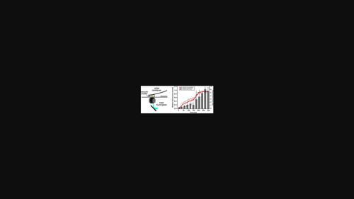
Vinculin is an integral component of integrin adhesions, where it functions as a molecular clutch coupling intracellular contraction to the extracellular matrix. Quantitating its contribution to the reinforcement of newly forming adhesions, however, requires ultrasensitive cell force assays covering short time and low force ranges. Here, we have combined atomic force microscopy-based single-cell force spectroscopy (SCFS) and optical tweezers force spectroscopy to investigate the role of vinculin in reinforcement of individual nascent adhesions during the first 5 min of cell contact with fibronectin or vitronectin. At minimal adhesion times (5-10 s), mouse embryonic fibroblast (MEF) wildtype (wt) and vinculin knock-out (vin(−/−)) cells develop comparable adhesion forces on the scale of several individual integrin-ligand bonds, confirming that vinculin is dispensable for adhesion initiation. In contrast, after 60 to 120 s, adhesion strength and traction reinforce quickly in wt cells, while remaining low in vin(−/−) cells. Re-expression of full-length vinculin or a constitutively active vinculin mutant (vinT12) in MEF vin(−/−) cells restored adhesion and traction with the same efficiency, while vinculin with a mutated talin-binding head region (vinA50I) or missing the actin-binding tail-domain (vin880) was ineffective. Integrating total internal reflection fluorescence imaging into the SCFS setup furthermore enabled us to correlate vinculin-green fluorescent protein (GFP) recruitment to nascent adhesion sites with the built-up of vinculin-dependent adhesion forces directly. Vinculin recruitment and cell adhesion reinforcement followed synchronous biphasic patterns, suggesting vinculin recruitment, but not activation, as the rate-limiting step for adhesion reinforcement. Combining sensitive SCFS with fluorescence microscopy thus provides insight into the temporal sequence of vinculin-dependent mechanical reinforcement in nascent integrin adhesions.
期刊介绍:
Journal of Molecular Recognition (JMR) publishes original research papers and reviews describing substantial advances in our understanding of molecular recognition phenomena in life sciences, covering all aspects from biochemistry, molecular biology, medicine, and biophysics. The research may employ experimental, theoretical and/or computational approaches.
The focus of the journal is on recognition phenomena involving biomolecules and their biological / biochemical partners rather than on the recognition of metal ions or inorganic compounds. Molecular recognition involves non-covalent specific interactions between two or more biological molecules, molecular aggregates, cellular modules or organelles, as exemplified by receptor-ligand, antigen-antibody, nucleic acid-protein, sugar-lectin, to mention just a few of the possible interactions. The journal invites manuscripts that aim to achieve a complete description of molecular recognition mechanisms between well-characterized biomolecules in terms of structure, dynamics and biological activity. Such studies may help the future development of new drugs and vaccines, although the experimental testing of new drugs and vaccines falls outside the scope of the journal. Manuscripts that describe the application of standard approaches and techniques to design or model new molecular entities or to describe interactions between biomolecules, but do not provide new insights into molecular recognition processes will not be considered. Similarly, manuscripts involving biomolecules uncharacterized at the sequence level (e.g. calf thymus DNA) will not be considered.
 求助内容:
求助内容: 应助结果提醒方式:


