{"title":"Bilateral duplicated hypoplastic superior cerebellar arteries one of which originates from a full-type fetal posterior cerebral artery.","authors":"Mohammed Assaad Alnafie","doi":"10.1007/s00276-023-03230-8","DOIUrl":null,"url":null,"abstract":"<p><strong>Purpose: </strong>The association of bilateral duplication of the superior cerebellar artery with an origin from the posterior cerebral artery is rare but of great interest to anatomists, radiologists, and surgeons. This article reports bilateral duplicated hypoplastic superior cerebellar arteries, one of which arises from a full-type fetal cerebral artery.</p><p><strong>Material and method: </strong>A 59-year-old woman admitted to the neurosurgery department for a subarachnoid hemorrhage underwent a brain CTA with 3D reconstruction using «3D slicer 4.11» software. Brain CTA and the 3D model were used to analyze the configuration of the posterior circulation.</p><p><strong>Results: </strong>CTA images and the 3D model showed an unusual configuration of the posterior circulation. The basilar artery prolonged the left vertebral artery, while the right vertebral artery ended in the right posterior inferior cerebellar artery. On both sides, a full-type fetal posterior cerebral artery and duplicated hypoplastic superior cerebellar artery were observed. Three cerebellar arteries arose from the basilar artery, while the fourth one emerged from the right fetal posterior cerebral artery.</p><p><strong>Conclusion: </strong>Knowledge of such a configuration of the posterior circulation and others is necessary before radiological and surgical procedures. It helps to understand hemodynamic events, and neurovascular conflicts, improve revascularization procedures, and avoid surgical arterial and nervous injuries.</p>","PeriodicalId":49296,"journal":{"name":"Surgical and Radiologic Anatomy","volume":" ","pages":"1295-1300"},"PeriodicalIF":1.2000,"publicationDate":"2023-10-01","publicationTypes":"Journal Article","fieldsOfStudy":null,"isOpenAccess":false,"openAccessPdf":"","citationCount":"0","resultStr":null,"platform":"Semanticscholar","paperid":null,"PeriodicalName":"Surgical and Radiologic Anatomy","FirstCategoryId":"3","ListUrlMain":"https://doi.org/10.1007/s00276-023-03230-8","RegionNum":4,"RegionCategory":"医学","ArticlePicture":[],"TitleCN":null,"AbstractTextCN":null,"PMCID":null,"EPubDate":"2023/8/10 0:00:00","PubModel":"Epub","JCR":"Q3","JCRName":"ANATOMY & MORPHOLOGY","Score":null,"Total":0}
引用次数: 0
Abstract
Purpose: The association of bilateral duplication of the superior cerebellar artery with an origin from the posterior cerebral artery is rare but of great interest to anatomists, radiologists, and surgeons. This article reports bilateral duplicated hypoplastic superior cerebellar arteries, one of which arises from a full-type fetal cerebral artery.
Material and method: A 59-year-old woman admitted to the neurosurgery department for a subarachnoid hemorrhage underwent a brain CTA with 3D reconstruction using «3D slicer 4.11» software. Brain CTA and the 3D model were used to analyze the configuration of the posterior circulation.
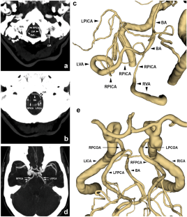
Results: CTA images and the 3D model showed an unusual configuration of the posterior circulation. The basilar artery prolonged the left vertebral artery, while the right vertebral artery ended in the right posterior inferior cerebellar artery. On both sides, a full-type fetal posterior cerebral artery and duplicated hypoplastic superior cerebellar artery were observed. Three cerebellar arteries arose from the basilar artery, while the fourth one emerged from the right fetal posterior cerebral artery.
Conclusion: Knowledge of such a configuration of the posterior circulation and others is necessary before radiological and surgical procedures. It helps to understand hemodynamic events, and neurovascular conflicts, improve revascularization procedures, and avoid surgical arterial and nervous injuries.
期刊介绍:
Anatomy is a morphological science which cannot fail to interest the clinician. The practical application of anatomical research to clinical problems necessitates special adaptation and selectivity in choosing from numerous international works. Although there is a tendency to believe that meaningful advances in anatomy are unlikely, constant revision is necessary. Surgical and Radiologic Anatomy, the first international journal of Clinical anatomy has been created in this spirit.
Its goal is to serve clinicians, regardless of speciality-physicians, surgeons, radiologists or other specialists-as an indispensable aid with which they can improve their knowledge of anatomy. Each issue includes: Original papers, review articles, articles on the anatomical bases of medical, surgical and radiological techniques, articles of normal radiologic anatomy, brief reviews of anatomical publications of clinical interest.
Particular attention is given to high quality illustrations, which are indispensable for a better understanding of anatomical problems.
Surgical and Radiologic Anatomy is a journal written by anatomists for clinicians with a special interest in anatomy.

 求助内容:
求助内容: 应助结果提醒方式:


