{"title":"A Case of Neurofibromatosis Type 1 Diagnosed after Idiopathic Rupture of Superficial Temporal Artery Pseudoaneurysm Requiring Endovascular Treatment.","authors":"Takashi Iwama, Katsuhiro Mizutani, Hajime Kubo, Masahiro Katsumata, Takenori Akiyama, Masahiro Toda","doi":"10.2176/jns-nmc.2022-0349","DOIUrl":null,"url":null,"abstract":"<p><p>Patients with neurofibromatosis type 1 not only have characteristic skin findings but are also known to have vascular disorders due to vascular vulnerability. A 44-year-old man with previously undiagnosed neurofibromatosis type 1 was brought to the emergency room due to a sudden subcutaneous hematoma with no history of trauma. Angiography revealed extravasation from the parietal branch of the right superficial temporal artery, which was embolized with n-butyl-2-cyanoacrylate. However, the next day, the patient exhibited an increased subcutaneous hematoma, and new extravascular leakage was detected at the frontal branch of the superficial temporal artery, which was also embolized with n-butyl-2-cyanoacrylate. The patient had physical findings characteristic of neurofibromatosis type 1, such as <i>café-au-lait</i> spots, and was subsequently diagnosed with neurofibromatosis type 1. No obvious neurofibroma or any other subcutaneous lesion associated with neurofibromatosis type 1 was identified in the affected area. Massive idiopathic arterial bleeding in the scalp, although infrequent, can be fatal. Neurofibromatosis type 1 should be considered when a subcutaneous scalp hematoma is observed without a history of trauma, even if the facial skin structure appears normal. Neurofibromatosis type 1 is also known to have multiple sources of hemorrhage. Thus, it is important to repeatedly evaluate vascular structures <i>via</i> cerebral angiography, contrast-enhanced computed tomography, and magnetic resonance imaging, if necessary.</p>","PeriodicalId":19260,"journal":{"name":"NMC Case Report Journal","volume":"10 ","pages":"125-130"},"PeriodicalIF":0.0000,"publicationDate":"2023-01-01","publicationTypes":"Journal Article","fieldsOfStudy":null,"isOpenAccess":false,"openAccessPdf":"https://ftp.ncbi.nlm.nih.gov/pub/pmc/oa_pdf/1f/29/2188-4226-10-0125.PMC10247217.pdf","citationCount":"1","resultStr":null,"platform":"Semanticscholar","paperid":null,"PeriodicalName":"NMC Case Report Journal","FirstCategoryId":"1085","ListUrlMain":"https://doi.org/10.2176/jns-nmc.2022-0349","RegionNum":0,"RegionCategory":null,"ArticlePicture":[],"TitleCN":null,"AbstractTextCN":null,"PMCID":null,"EPubDate":"","PubModel":"","JCR":"","JCRName":"","Score":null,"Total":0}
引用次数: 1
Abstract
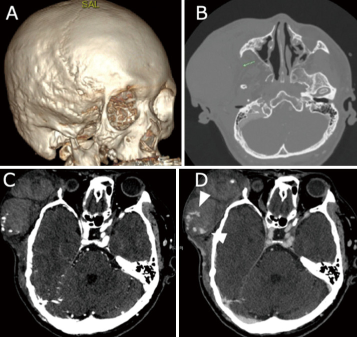
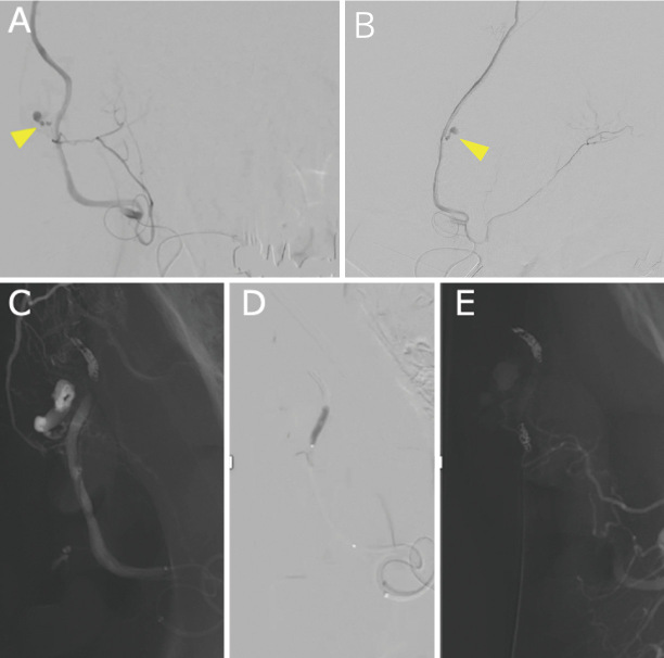
Patients with neurofibromatosis type 1 not only have characteristic skin findings but are also known to have vascular disorders due to vascular vulnerability. A 44-year-old man with previously undiagnosed neurofibromatosis type 1 was brought to the emergency room due to a sudden subcutaneous hematoma with no history of trauma. Angiography revealed extravasation from the parietal branch of the right superficial temporal artery, which was embolized with n-butyl-2-cyanoacrylate. However, the next day, the patient exhibited an increased subcutaneous hematoma, and new extravascular leakage was detected at the frontal branch of the superficial temporal artery, which was also embolized with n-butyl-2-cyanoacrylate. The patient had physical findings characteristic of neurofibromatosis type 1, such as café-au-lait spots, and was subsequently diagnosed with neurofibromatosis type 1. No obvious neurofibroma or any other subcutaneous lesion associated with neurofibromatosis type 1 was identified in the affected area. Massive idiopathic arterial bleeding in the scalp, although infrequent, can be fatal. Neurofibromatosis type 1 should be considered when a subcutaneous scalp hematoma is observed without a history of trauma, even if the facial skin structure appears normal. Neurofibromatosis type 1 is also known to have multiple sources of hemorrhage. Thus, it is important to repeatedly evaluate vascular structures via cerebral angiography, contrast-enhanced computed tomography, and magnetic resonance imaging, if necessary.


 求助内容:
求助内容: 应助结果提醒方式:


