Anni Gålne, Olof Enqvist, Anna Sundlöv, Kristian Valind, David Minarik, Elin Trägårdh
{"title":"AI-based quantification of whole-body tumour burden on somatostatin receptor PET/CT.","authors":"Anni Gålne, Olof Enqvist, Anna Sundlöv, Kristian Valind, David Minarik, Elin Trägårdh","doi":"10.1186/s41824-023-00172-7","DOIUrl":null,"url":null,"abstract":"<p><strong>Background: </strong>Segmenting the whole-body somatostatin receptor-expressing tumour volume (SRETVwb) on positron emission tomography/computed tomography (PET/CT) images is highly time-consuming but has shown value as an independent prognostic factor for survival. An automatic method to measure SRETVwb could improve disease status assessment and provide a tool for prognostication. This study aimed to develop an artificial intelligence (AI)-based method to detect and quantify SRETVwb and total lesion somatostatin receptor expression (TLSREwb) from [<sup>68</sup>Ga]Ga-DOTA-TOC/TATE PET/CT images.</p><p><strong>Methods: </strong>A UNet3D convolutional neural network (CNN) was used to train an AI model with [<sup>68</sup>Ga]Ga-DOTA-TOC/TATE PET/CT images, where all tumours were manually segmented with a semi-automatic method. The training set consisted of 148 patients, of which 108 had PET-positive tumours. The test group consisted of 30 patients, of which 25 had PET-positive tumours. Two physicians segmented tumours in the test group for comparison with the AI model.</p><p><strong>Results: </strong>There were good correlations between the segmented SRETVwb and TLSREwb by the AI model and the physicians, with Spearman rank correlation coefficients of r = 0.78 and r = 0.73, respectively, for SRETVwb and r = 0.83 and r = 0.81, respectively, for TLSREwb. The sensitivity on a lesion detection level was 80% and 79%, and the positive predictive value was 83% and 84% when comparing the AI model with the two physicians.</p><p><strong>Conclusion: </strong>It was possible to develop an AI model to segment SRETVwb and TLSREwb with high performance. A fully automated method makes quantification of tumour burden achievable and has the potential to be more widely used when assessing PET/CT images.</p>","PeriodicalId":36160,"journal":{"name":"European Journal of Hybrid Imaging","volume":"7 1","pages":"14"},"PeriodicalIF":1.2000,"publicationDate":"2023-08-07","publicationTypes":"Journal Article","fieldsOfStudy":null,"isOpenAccess":false,"openAccessPdf":"https://www.ncbi.nlm.nih.gov/pmc/articles/PMC10404578/pdf/","citationCount":"0","resultStr":null,"platform":"Semanticscholar","paperid":null,"PeriodicalName":"European Journal of Hybrid Imaging","FirstCategoryId":"1085","ListUrlMain":"https://doi.org/10.1186/s41824-023-00172-7","RegionNum":0,"RegionCategory":null,"ArticlePicture":[],"TitleCN":null,"AbstractTextCN":null,"PMCID":null,"EPubDate":"","PubModel":"","JCR":"Q3","JCRName":"RADIOLOGY, NUCLEAR MEDICINE & MEDICAL IMAGING","Score":null,"Total":0}
引用次数: 0
Abstract
Background: Segmenting the whole-body somatostatin receptor-expressing tumour volume (SRETVwb) on positron emission tomography/computed tomography (PET/CT) images is highly time-consuming but has shown value as an independent prognostic factor for survival. An automatic method to measure SRETVwb could improve disease status assessment and provide a tool for prognostication. This study aimed to develop an artificial intelligence (AI)-based method to detect and quantify SRETVwb and total lesion somatostatin receptor expression (TLSREwb) from [68Ga]Ga-DOTA-TOC/TATE PET/CT images.
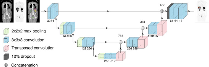
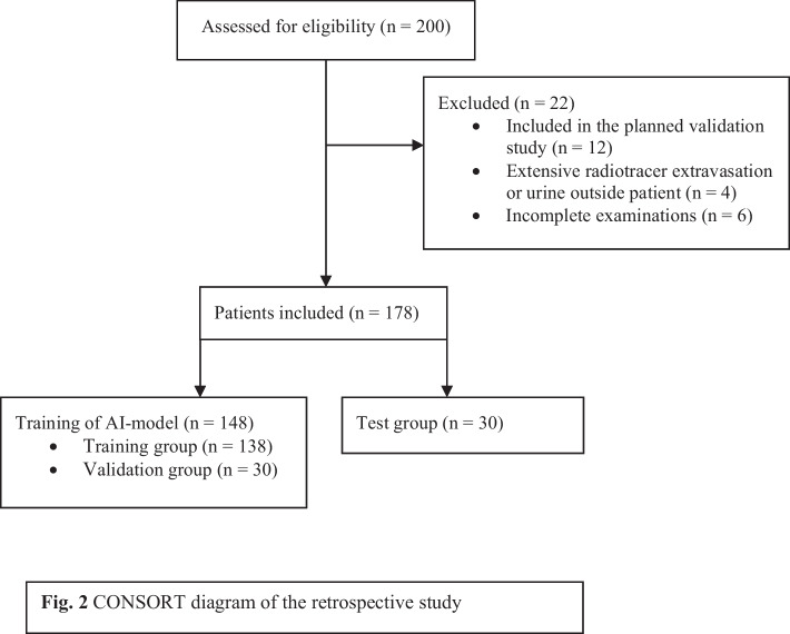
Methods: A UNet3D convolutional neural network (CNN) was used to train an AI model with [68Ga]Ga-DOTA-TOC/TATE PET/CT images, where all tumours were manually segmented with a semi-automatic method. The training set consisted of 148 patients, of which 108 had PET-positive tumours. The test group consisted of 30 patients, of which 25 had PET-positive tumours. Two physicians segmented tumours in the test group for comparison with the AI model.
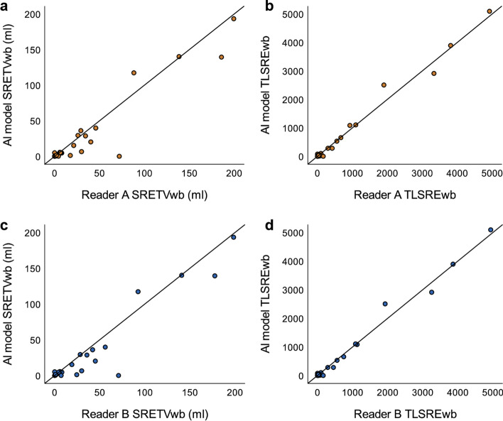
Results: There were good correlations between the segmented SRETVwb and TLSREwb by the AI model and the physicians, with Spearman rank correlation coefficients of r = 0.78 and r = 0.73, respectively, for SRETVwb and r = 0.83 and r = 0.81, respectively, for TLSREwb. The sensitivity on a lesion detection level was 80% and 79%, and the positive predictive value was 83% and 84% when comparing the AI model with the two physicians.
Conclusion: It was possible to develop an AI model to segment SRETVwb and TLSREwb with high performance. A fully automated method makes quantification of tumour burden achievable and has the potential to be more widely used when assessing PET/CT images.

 求助内容:
求助内容: 应助结果提醒方式:


