Markus E Krogager, Rasmus H Dahl, Lars Poulsgaard, Kåre Fugleholm, Tom Sehested, Ronni Mikkelsen, Jørgen Tranum-Jensen, Tiit I Mathiesen, Goetz Benndorf
{"title":"Combined cone-beam CT imaging and microsurgical dissection of cadaver specimens to study cerebral venous anatomy: a technical note.","authors":"Markus E Krogager, Rasmus H Dahl, Lars Poulsgaard, Kåre Fugleholm, Tom Sehested, Ronni Mikkelsen, Jørgen Tranum-Jensen, Tiit I Mathiesen, Goetz Benndorf","doi":"10.1007/s00276-023-03195-8","DOIUrl":null,"url":null,"abstract":"<p><strong>Purpose: </strong>Cadaver dissections and X-ray based 3D angiography are considered gold standards for studying neurovascular anatomy. We sought to develop a model that utilize the combination of both these techniques to improve current tools for anatomical research, teaching and preoperative surgical planning, particularly addressing the venous system of the brain.</p><p><strong>Materials and methods: </strong>Seven ethanol-fixed human cadaveric heads and one arm were injected with a latex-barium mixture into the internal jugular veins and the brachial artery. After the ethanol-based fixation, specimens were scanned by high-resolution cone-beam CT and images were post-processed on a 3D-workstation. Subsequent, microsurgical dissections were performed by an experienced neurosurgeon and venous anatomy was compared with relevant 3D venograms.</p><p><strong>Results: </strong>Latex-barium mixtures resulted in a homogenous cast with filling of the cerebral venous structures down to 150 μm in diameter. The ethanol-based preparation of the cadaveric brains allowed for near-realistic microsurgical maneuverability during dissection. The model improves assessment of the venous system for anatomical education and hands-on surgical training.</p><p><strong>Conclusion: </strong>To our knowledge we describe the first preparation method which combines near-realistic microsurgical dissection of human heads with high-resolution 3D imaging of the cerebral venous system in the same specimens.</p>","PeriodicalId":49296,"journal":{"name":"Surgical and Radiologic Anatomy","volume":" ","pages":"1177-1184"},"PeriodicalIF":1.2000,"publicationDate":"2023-09-01","publicationTypes":"Journal Article","fieldsOfStudy":null,"isOpenAccess":false,"openAccessPdf":"https://www.ncbi.nlm.nih.gov/pmc/articles/PMC10514096/pdf/","citationCount":"0","resultStr":null,"platform":"Semanticscholar","paperid":null,"PeriodicalName":"Surgical and Radiologic Anatomy","FirstCategoryId":"3","ListUrlMain":"https://doi.org/10.1007/s00276-023-03195-8","RegionNum":4,"RegionCategory":"医学","ArticlePicture":[],"TitleCN":null,"AbstractTextCN":null,"PMCID":null,"EPubDate":"2023/8/5 0:00:00","PubModel":"Epub","JCR":"Q3","JCRName":"ANATOMY & MORPHOLOGY","Score":null,"Total":0}
引用次数: 0
Abstract
Purpose: Cadaver dissections and X-ray based 3D angiography are considered gold standards for studying neurovascular anatomy. We sought to develop a model that utilize the combination of both these techniques to improve current tools for anatomical research, teaching and preoperative surgical planning, particularly addressing the venous system of the brain.
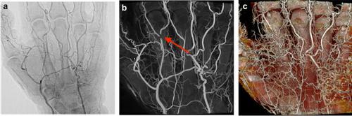
Materials and methods: Seven ethanol-fixed human cadaveric heads and one arm were injected with a latex-barium mixture into the internal jugular veins and the brachial artery. After the ethanol-based fixation, specimens were scanned by high-resolution cone-beam CT and images were post-processed on a 3D-workstation. Subsequent, microsurgical dissections were performed by an experienced neurosurgeon and venous anatomy was compared with relevant 3D venograms.
Results: Latex-barium mixtures resulted in a homogenous cast with filling of the cerebral venous structures down to 150 μm in diameter. The ethanol-based preparation of the cadaveric brains allowed for near-realistic microsurgical maneuverability during dissection. The model improves assessment of the venous system for anatomical education and hands-on surgical training.
Conclusion: To our knowledge we describe the first preparation method which combines near-realistic microsurgical dissection of human heads with high-resolution 3D imaging of the cerebral venous system in the same specimens.
期刊介绍:
Anatomy is a morphological science which cannot fail to interest the clinician. The practical application of anatomical research to clinical problems necessitates special adaptation and selectivity in choosing from numerous international works. Although there is a tendency to believe that meaningful advances in anatomy are unlikely, constant revision is necessary. Surgical and Radiologic Anatomy, the first international journal of Clinical anatomy has been created in this spirit.
Its goal is to serve clinicians, regardless of speciality-physicians, surgeons, radiologists or other specialists-as an indispensable aid with which they can improve their knowledge of anatomy. Each issue includes: Original papers, review articles, articles on the anatomical bases of medical, surgical and radiological techniques, articles of normal radiologic anatomy, brief reviews of anatomical publications of clinical interest.
Particular attention is given to high quality illustrations, which are indispensable for a better understanding of anatomical problems.
Surgical and Radiologic Anatomy is a journal written by anatomists for clinicians with a special interest in anatomy.

 求助内容:
求助内容: 应助结果提醒方式:


