Eva Liu, Pramath Kakodkar, Henry Pan, Amy Zhou, Patrick Toyota, Amit Rahul Persad, Kristen Marciniuk, Chunjie Wang, Roland Nikolaus Auer, Stephen Sanche, Aleksander Vitali, Julia Radic
{"title":"Pediatric intracranial tuberculoma: illustrative case.","authors":"Eva Liu, Pramath Kakodkar, Henry Pan, Amy Zhou, Patrick Toyota, Amit Rahul Persad, Kristen Marciniuk, Chunjie Wang, Roland Nikolaus Auer, Stephen Sanche, Aleksander Vitali, Julia Radic","doi":"10.3171/CASE23236","DOIUrl":null,"url":null,"abstract":"<p><strong>Background: </strong>Tuberculosis is an airborne disease caused by Mycobacterium tuberculosis. Intracranial tuberculoma is a rare complication of extrapulmonary tuberculosis due to hematogenous spread to subpial and subependymal regions. Intracranial tuberculoma can occur with or without meningitis.</p><p><strong>Observations: </strong>A 3-year-old male who had recently emigrated from Sudan presented to the emergency department with right-sided seizures lasting 30 minutes, which were aborted with levetiracetam and midazolam. Head computed tomography revealed a multilobulated left supratentorial mass with solid and cystic components and measuring 8.0 × 4.8 × 6.5 cm. The patient had successful resection of the mass, which was positive for M. tuberculosis. He was started on rifampin, isoniazid, pyrazinamide, ethambutol, and fluoroquinolone and was discharged home in stable condition.</p><p><strong>Lessons: </strong>A literature review on pediatric intracranial tuberculoma was performed, which included 48 studies (n = 49). The mean age was 8.8 ± 5.4 years with a slight female predilection (59%). Predominant solitary tuberculomas (63%) were preferentially managed with both resection and antituberculosis therapy (ATT), whereas multifocal tuberculomas were preferentially managed with ATT. Intracranial tuberculoma is a rare but treatable cause of space-occupying lesions in children. Clinicians should maintain a high level of suspicion in patients from endemic regions and involve the infectious disease service early.</p>","PeriodicalId":16554,"journal":{"name":"Journal of Neurosurgery: Case Lessons","volume":"6 4","pages":""},"PeriodicalIF":0.0000,"publicationDate":"2023-07-24","publicationTypes":"Journal Article","fieldsOfStudy":null,"isOpenAccess":false,"openAccessPdf":"https://ftp.ncbi.nlm.nih.gov/pub/pmc/oa_pdf/bb/12/CASE23236.PMC10555599.pdf","citationCount":"0","resultStr":null,"platform":"Semanticscholar","paperid":null,"PeriodicalName":"Journal of Neurosurgery: Case Lessons","FirstCategoryId":"1085","ListUrlMain":"https://doi.org/10.3171/CASE23236","RegionNum":0,"RegionCategory":null,"ArticlePicture":[],"TitleCN":null,"AbstractTextCN":null,"PMCID":null,"EPubDate":"","PubModel":"","JCR":"","JCRName":"","Score":null,"Total":0}
引用次数: 0
Abstract
Background: Tuberculosis is an airborne disease caused by Mycobacterium tuberculosis. Intracranial tuberculoma is a rare complication of extrapulmonary tuberculosis due to hematogenous spread to subpial and subependymal regions. Intracranial tuberculoma can occur with or without meningitis.
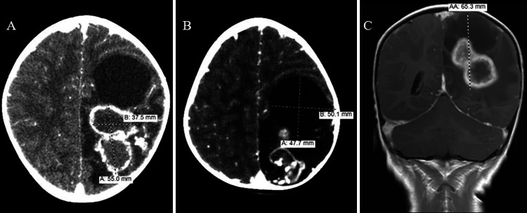
Observations: A 3-year-old male who had recently emigrated from Sudan presented to the emergency department with right-sided seizures lasting 30 minutes, which were aborted with levetiracetam and midazolam. Head computed tomography revealed a multilobulated left supratentorial mass with solid and cystic components and measuring 8.0 × 4.8 × 6.5 cm. The patient had successful resection of the mass, which was positive for M. tuberculosis. He was started on rifampin, isoniazid, pyrazinamide, ethambutol, and fluoroquinolone and was discharged home in stable condition.
Lessons: A literature review on pediatric intracranial tuberculoma was performed, which included 48 studies (n = 49). The mean age was 8.8 ± 5.4 years with a slight female predilection (59%). Predominant solitary tuberculomas (63%) were preferentially managed with both resection and antituberculosis therapy (ATT), whereas multifocal tuberculomas were preferentially managed with ATT. Intracranial tuberculoma is a rare but treatable cause of space-occupying lesions in children. Clinicians should maintain a high level of suspicion in patients from endemic regions and involve the infectious disease service early.

 求助内容:
求助内容: 应助结果提醒方式:


