{"title":"Pediatric internal auditory canal cavernous hemangioma with rapid progression of sensorineural hearing loss: illustrative case.","authors":"Hiroshi Hyakusoku, Yoshihide Tanaka, Yusuke Tsuchiya, Meijin Nakayama","doi":"10.3171/CASE23141","DOIUrl":null,"url":null,"abstract":"<p><strong>Background: </strong>Cavernous hemangioma of the internal auditory canal is extremely rare and is characterized by symptoms such as vertigo, sensorineural hearing loss, and facial nerve dysfunction.</p><p><strong>Observations: </strong>A health examination on an 11-year-old female in the fifth grade revealed hearing loss in the left ear. She also had dizziness that had persisted for approximately 1 year. Pure-tone audiometry revealed sensorineural hearing loss in her left ear. Rightward horizontal and rotatory nystagmus was detected. Facial paralysis was not present. Magnetic resonance imaging showed a lesion that was suspected to be hemangioma. The authors selected a left suboccipital retrosigmoid approach. The tumor showed a berry-tufted appearance throughout the cerebellopontine angle. The seventh cranial nerve penetrated the tumor and partly circulated outside the tumor with marked adhesion. The authors partially resected the tumor to avoid damaging the facial nerve. A histological examination identified cavernous hemangioma.</p><p><strong>Lessons: </strong>The fundamental treatment for cavernous hemangioma of the internal auditory canal is complete surgical removal; however, any surgical intervention may result in hearing loss and facial paralysis. The extent of surgery needs to be decided intraoperatively based on the balance between preoperative symptoms and postoperative complications.</p>","PeriodicalId":16554,"journal":{"name":"Journal of Neurosurgery: Case Lessons","volume":"5 22","pages":""},"PeriodicalIF":0.0000,"publicationDate":"2023-05-29","publicationTypes":"Journal Article","fieldsOfStudy":null,"isOpenAccess":false,"openAccessPdf":"https://ftp.ncbi.nlm.nih.gov/pub/pmc/oa_pdf/da/9f/CASE23141.PMC10550673.pdf","citationCount":"0","resultStr":null,"platform":"Semanticscholar","paperid":null,"PeriodicalName":"Journal of Neurosurgery: Case Lessons","FirstCategoryId":"1085","ListUrlMain":"https://doi.org/10.3171/CASE23141","RegionNum":0,"RegionCategory":null,"ArticlePicture":[],"TitleCN":null,"AbstractTextCN":null,"PMCID":null,"EPubDate":"","PubModel":"","JCR":"","JCRName":"","Score":null,"Total":0}
引用次数: 0
Abstract
Background: Cavernous hemangioma of the internal auditory canal is extremely rare and is characterized by symptoms such as vertigo, sensorineural hearing loss, and facial nerve dysfunction.
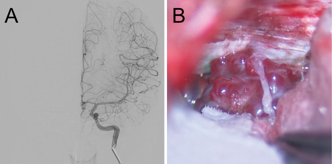
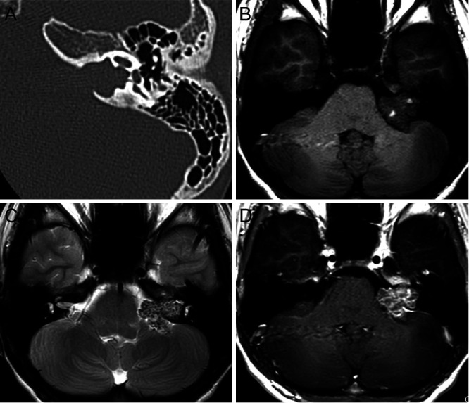
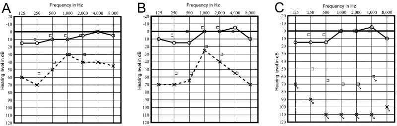
Observations: A health examination on an 11-year-old female in the fifth grade revealed hearing loss in the left ear. She also had dizziness that had persisted for approximately 1 year. Pure-tone audiometry revealed sensorineural hearing loss in her left ear. Rightward horizontal and rotatory nystagmus was detected. Facial paralysis was not present. Magnetic resonance imaging showed a lesion that was suspected to be hemangioma. The authors selected a left suboccipital retrosigmoid approach. The tumor showed a berry-tufted appearance throughout the cerebellopontine angle. The seventh cranial nerve penetrated the tumor and partly circulated outside the tumor with marked adhesion. The authors partially resected the tumor to avoid damaging the facial nerve. A histological examination identified cavernous hemangioma.
Lessons: The fundamental treatment for cavernous hemangioma of the internal auditory canal is complete surgical removal; however, any surgical intervention may result in hearing loss and facial paralysis. The extent of surgery needs to be decided intraoperatively based on the balance between preoperative symptoms and postoperative complications.

 求助内容:
求助内容: 应助结果提醒方式:


