Héloïse Lebas, Alexandre Boutigny, Clémence Maupu, Jonas Salfati, Cyrille Orset, Mikael Mazighi, Philippe Bonnin, Yacine Boulaftali
{"title":"Imaging Cerebral Arteries Tortuosity and Velocities by Transcranial Doppler Ultrasound Is a Reliable Assessment of Brain Aneurysm in Mouse Models.","authors":"Héloïse Lebas, Alexandre Boutigny, Clémence Maupu, Jonas Salfati, Cyrille Orset, Mikael Mazighi, Philippe Bonnin, Yacine Boulaftali","doi":"10.1161/SVIN.122.000476","DOIUrl":null,"url":null,"abstract":"<p><strong>Background: </strong>During the past few decades, several pathophysiological processes contributing to intracranial aneurysm (IA) rupture have been identified, including irregular IA shape, altered hemodynamic stress within the IA, and vessel wall inflammation. The use of preclinical models of IA and imaging tools is paramount to better understand the underlying disease mechanisms.</p><p><strong>Methods: </strong>We used 2 established mouse models of IA, and we analyzed the progression of the IA by magnetic resonance imaging, transcranial Doppler, and histology.</p><p><strong>Results: </strong>In both models of IA, we observed, by transcranial Doppler, a significant decrease of the blood velocities and wall shear stress of the internal carotid arteries. We also observed the formation of tortuous arteries in both models that were correlated with the presence of an aneurysm as confirmed by magnetic resonance imaging and histology. A high grade of tortuosity is associated with a significant decrease of the mean blood flow velocities and a greater artery dilation.</p><p><strong>Conclusions: </strong>Transcranial Doppler is a robust and convenient imaging method to evaluate the progression of IA. Detection of decreased blood flow velocities and increased tortuosity can be used as reliable indicators of IA.</p>","PeriodicalId":74875,"journal":{"name":"Stroke (Hoboken, N.J.)","volume":"3 2","pages":"e000476"},"PeriodicalIF":2.8000,"publicationDate":"2023-03-07","publicationTypes":"Journal Article","fieldsOfStudy":null,"isOpenAccess":false,"openAccessPdf":"https://ftp.ncbi.nlm.nih.gov/pub/pmc/oa_pdf/dd/a8/SVI2-3-e000476.PMC10368188.pdf","citationCount":"0","resultStr":null,"platform":"Semanticscholar","paperid":null,"PeriodicalName":"Stroke (Hoboken, N.J.)","FirstCategoryId":"1085","ListUrlMain":"https://doi.org/10.1161/SVIN.122.000476","RegionNum":0,"RegionCategory":null,"ArticlePicture":[],"TitleCN":null,"AbstractTextCN":null,"PMCID":null,"EPubDate":"","PubModel":"","JCR":"Q3","JCRName":"CLINICAL NEUROLOGY","Score":null,"Total":0}
引用次数: 0
Abstract
Background: During the past few decades, several pathophysiological processes contributing to intracranial aneurysm (IA) rupture have been identified, including irregular IA shape, altered hemodynamic stress within the IA, and vessel wall inflammation. The use of preclinical models of IA and imaging tools is paramount to better understand the underlying disease mechanisms.
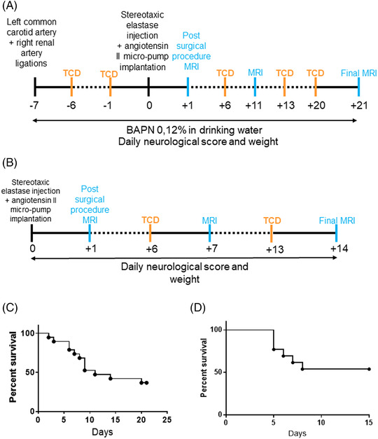
Methods: We used 2 established mouse models of IA, and we analyzed the progression of the IA by magnetic resonance imaging, transcranial Doppler, and histology.
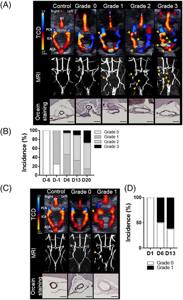
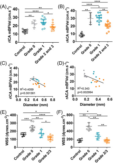
Results: In both models of IA, we observed, by transcranial Doppler, a significant decrease of the blood velocities and wall shear stress of the internal carotid arteries. We also observed the formation of tortuous arteries in both models that were correlated with the presence of an aneurysm as confirmed by magnetic resonance imaging and histology. A high grade of tortuosity is associated with a significant decrease of the mean blood flow velocities and a greater artery dilation.
Conclusions: Transcranial Doppler is a robust and convenient imaging method to evaluate the progression of IA. Detection of decreased blood flow velocities and increased tortuosity can be used as reliable indicators of IA.

 求助内容:
求助内容: 应助结果提醒方式:


