{"title":"Imaging Characteristics of Breast Lymphoma; a Case Series.","authors":"Sara Rehman, Muhammad Atif Naveed, Javaria Aleem","doi":"10.37029/jcas.v6i1.305","DOIUrl":null,"url":null,"abstract":"<p><strong>Introduction: </strong>Breast involvement by lymphoma is rare. It can occur as a primary breast tumour or as an extranodal manifestation of the systemic disease. The imaging features of breast lymphoma (BL) are not characteristic. Biopsy is necessary for diagnosis due to non-specific imaging features.</p><p><strong>Materials and methods: </strong>A retrospective electronic medical chart review was conducted of patients diagnosed with lymphoma of breast that underwent diagnostic radiological procedures (including mammography, ultrasound breast, computed tomography (CT) scan and positron emission tomography (PET/CT) scan from 1 July 2018 to 31 March 2019 at Shaukat Khanum Memorial Cancer Hospital and Research Centre, Pakistan.</p><p><strong>Results: </strong>Four patients were identified. On mammogram, the most common finding consisted of the presence of high-density masses with circumscribed or indistinct margins. On ultrasound, hypoechoic masses and indistinct diffuse infiltrative patterns were observed. PET/CT and CT were helpful in detecting extramammary sites of disease and for classifying the disease into primary or secondary BL.</p><p><strong>Conclusion: </strong>The early diagnosis of the BL is important. The radiologists should include lymphoma in the differential diagnosis when there is the absence of microcalcifications or spiculated margins on mammography and ultrasound.</p>","PeriodicalId":73631,"journal":{"name":"Journal of cancer & allied specialties","volume":"6 1","pages":"e305"},"PeriodicalIF":0.0000,"publicationDate":"2020-01-06","publicationTypes":"Journal Article","fieldsOfStudy":null,"isOpenAccess":false,"openAccessPdf":"https://ftp.ncbi.nlm.nih.gov/pub/pmc/oa_pdf/af/aa/JCAS-6-305.PMC10166316.pdf","citationCount":"0","resultStr":null,"platform":"Semanticscholar","paperid":null,"PeriodicalName":"Journal of cancer & allied specialties","FirstCategoryId":"1085","ListUrlMain":"https://doi.org/10.37029/jcas.v6i1.305","RegionNum":0,"RegionCategory":null,"ArticlePicture":[],"TitleCN":null,"AbstractTextCN":null,"PMCID":null,"EPubDate":"2020/1/1 0:00:00","PubModel":"eCollection","JCR":"","JCRName":"","Score":null,"Total":0}
引用次数: 0
Abstract
Introduction: Breast involvement by lymphoma is rare. It can occur as a primary breast tumour or as an extranodal manifestation of the systemic disease. The imaging features of breast lymphoma (BL) are not characteristic. Biopsy is necessary for diagnosis due to non-specific imaging features.
Materials and methods: A retrospective electronic medical chart review was conducted of patients diagnosed with lymphoma of breast that underwent diagnostic radiological procedures (including mammography, ultrasound breast, computed tomography (CT) scan and positron emission tomography (PET/CT) scan from 1 July 2018 to 31 March 2019 at Shaukat Khanum Memorial Cancer Hospital and Research Centre, Pakistan.
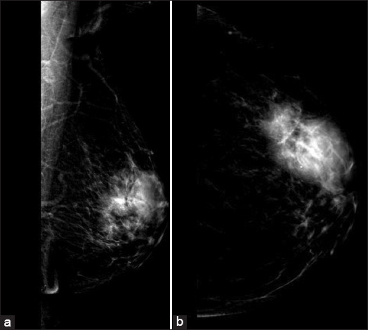
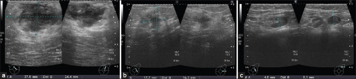
Results: Four patients were identified. On mammogram, the most common finding consisted of the presence of high-density masses with circumscribed or indistinct margins. On ultrasound, hypoechoic masses and indistinct diffuse infiltrative patterns were observed. PET/CT and CT were helpful in detecting extramammary sites of disease and for classifying the disease into primary or secondary BL.
Conclusion: The early diagnosis of the BL is important. The radiologists should include lymphoma in the differential diagnosis when there is the absence of microcalcifications or spiculated margins on mammography and ultrasound.

 求助内容:
求助内容: 应助结果提醒方式:


