A Daniel Davidar, Brendan F Judy, Andrew M Hersh, Carly Weber-Levine, Safwan Alomari, Arjun K Menta, Kelly Jiang, Meghana Bhimreddy, Mir Hussain, Neil R Crawford, Majid Khan, Gary Gong, Nicholas Theodore
{"title":"Robot-assisted screw fixation in a cadaver utilizing magnetic resonance imaging-based synthetic computed tomography: toward radiation-free spine surgery. Illustrative case.","authors":"A Daniel Davidar, Brendan F Judy, Andrew M Hersh, Carly Weber-Levine, Safwan Alomari, Arjun K Menta, Kelly Jiang, Meghana Bhimreddy, Mir Hussain, Neil R Crawford, Majid Khan, Gary Gong, Nicholas Theodore","doi":"10.3171/CASE23120","DOIUrl":null,"url":null,"abstract":"<p><strong>Background: </strong>Synthetic computed tomography (sCT) can be created from magnetic resonance imaging (MRI) utilizing newer software. sCT is yet to be explored as a possible alternative to routine CT (rCT). In this study, rCT scans and MRI-derived sCT scans were obtained on a cadaver. Morphometric analysis was performed comparing the 2 scans. The ExcelsiusGPS robot was used to place lumbosacral screws with both rCT and sCT images.</p><p><strong>Observations: </strong>In total, 14 screws were placed. All screws were grade A on the Gertzbein-Robbins scale. The mean surface distance difference between rCT and sCT on a reconstructed software model was -0.02 ± 0.05 mm, the mean absolute surface distance was 0.24 ± 0.05 mm, and the mean absolute error of radiodensity was 92.88 ± 10.53 HU. The overall mean tip distance for the sCT versus rCT was 1.74 ± 1.1 versus 2.36 ± 1.6 mm (p = 0.24); mean tail distance for the sCT versus rCT was 1.93 ± 0.88 versus 2.81 ± 1.03 mm (p = 0.07); and mean angular deviation for the sCT versus rCT was 3.2° ± 2.05° versus 4.04°± 2.71° (p = 0.53).</p><p><strong>Lessons: </strong>MRI-based sCT yielded results comparable to those of rCT in both morphometric analysis and robot-assisted lumbosacral screw placement in a cadaver study.</p>","PeriodicalId":16554,"journal":{"name":"Journal of Neurosurgery: Case Lessons","volume":"6 2","pages":""},"PeriodicalIF":0.0000,"publicationDate":"2023-07-10","publicationTypes":"Journal Article","fieldsOfStudy":null,"isOpenAccess":false,"openAccessPdf":"https://ftp.ncbi.nlm.nih.gov/pub/pmc/oa_pdf/49/c3/CASE23120.PMC10555644.pdf","citationCount":"1","resultStr":null,"platform":"Semanticscholar","paperid":null,"PeriodicalName":"Journal of Neurosurgery: Case Lessons","FirstCategoryId":"1085","ListUrlMain":"https://doi.org/10.3171/CASE23120","RegionNum":0,"RegionCategory":null,"ArticlePicture":[],"TitleCN":null,"AbstractTextCN":null,"PMCID":null,"EPubDate":"","PubModel":"","JCR":"","JCRName":"","Score":null,"Total":0}
引用次数: 1
Abstract
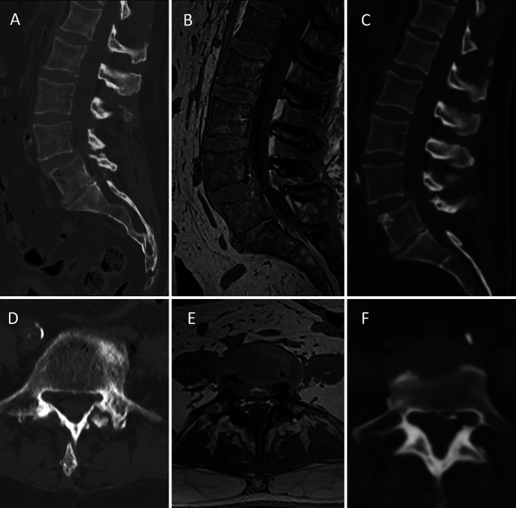
Background: Synthetic computed tomography (sCT) can be created from magnetic resonance imaging (MRI) utilizing newer software. sCT is yet to be explored as a possible alternative to routine CT (rCT). In this study, rCT scans and MRI-derived sCT scans were obtained on a cadaver. Morphometric analysis was performed comparing the 2 scans. The ExcelsiusGPS robot was used to place lumbosacral screws with both rCT and sCT images.
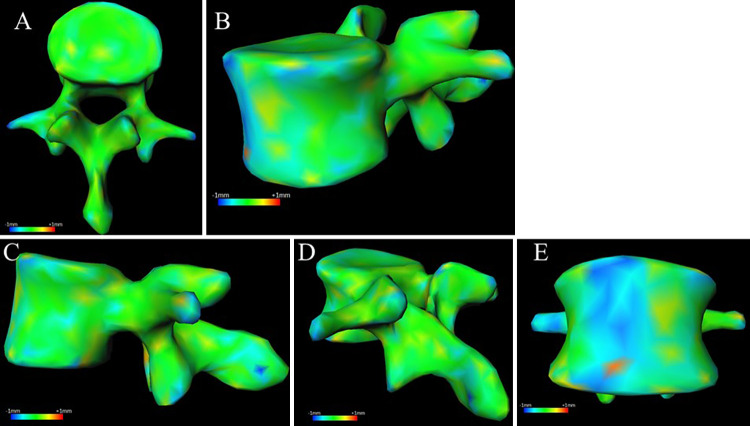
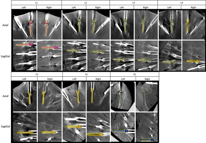
Observations: In total, 14 screws were placed. All screws were grade A on the Gertzbein-Robbins scale. The mean surface distance difference between rCT and sCT on a reconstructed software model was -0.02 ± 0.05 mm, the mean absolute surface distance was 0.24 ± 0.05 mm, and the mean absolute error of radiodensity was 92.88 ± 10.53 HU. The overall mean tip distance for the sCT versus rCT was 1.74 ± 1.1 versus 2.36 ± 1.6 mm (p = 0.24); mean tail distance for the sCT versus rCT was 1.93 ± 0.88 versus 2.81 ± 1.03 mm (p = 0.07); and mean angular deviation for the sCT versus rCT was 3.2° ± 2.05° versus 4.04°± 2.71° (p = 0.53).
Lessons: MRI-based sCT yielded results comparable to those of rCT in both morphometric analysis and robot-assisted lumbosacral screw placement in a cadaver study.

 求助内容:
求助内容: 应助结果提醒方式:


