In Tae Moon, Sun-Hwa Kim, Jung Yeon Chin, Sung Hun Park, Chang-Hwan Yoon, Tae-Jin Youn, In-Ho Chae, Si-Hyuck Kang
{"title":"Accuracy of Artificial Intelligence-Based Automated Quantitative Coronary Angiography Compared to Intravascular Ultrasound: Retrospective Cohort Study.","authors":"In Tae Moon, Sun-Hwa Kim, Jung Yeon Chin, Sung Hun Park, Chang-Hwan Yoon, Tae-Jin Youn, In-Ho Chae, Si-Hyuck Kang","doi":"10.2196/45299","DOIUrl":null,"url":null,"abstract":"<p><strong>Background: </strong>An accurate quantitative analysis of coronary artery stenotic lesions is essential to make optimal clinical decisions. Recent advances in computer vision and machine learning technology have enabled the automated analysis of coronary angiography.</p><p><strong>Objective: </strong>The aim of this paper is to validate the performance of artificial intelligence-based quantitative coronary angiography (AI-QCA) in comparison with that of intravascular ultrasound (IVUS).</p><p><strong>Methods: </strong>This retrospective study included patients who underwent IVUS-guided coronary intervention at a single tertiary center in Korea. Proximal and distal reference areas, minimal luminal area, percent plaque burden, and lesion length were measured by AI-QCA and human experts using IVUS. First, fully automated QCA analysis was compared with IVUS analysis. Next, we adjusted the proximal and distal margins of AI-QCA to avoid geographic mismatch. Scatter plots, Pearson correlation coefficients, and Bland-Altman were used to analyze the data.</p><p><strong>Results: </strong>A total of 54 significant lesions were analyzed in 47 patients. The proximal and distal reference areas, as well as the minimal luminal area, showed moderate to strong correlation between the 2 modalities (correlation coefficients of 0.57, 0.80, and 0.52, respectively; P<.001). The correlation was weaker for percent area stenosis and lesion length, although statistically significant (correlation coefficients of 0.29 and 0.33, respectively). AI-QCA tended to measure reference vessel areas smaller and lesion lengths shorter than IVUS did. Systemic proportional bias was not observed in Bland-Altman plots. The biggest cause of bias originated from the geographic mismatch of AI-QCA with IVUS. Discrepancies in the proximal or distal lesion margins were observed between the 2 modalities, which were more frequent at the distal margins. After the adjustment of proximal or distal margins, there was a stronger correlation of proximal and distal reference areas between AI-QCA and IVUS (correlation coefficients of 0.70 and 0.83, respectively).</p><p><strong>Conclusions: </strong>AI-QCA showed a moderate to strong correlation compared with IVUS in analyzing coronary lesions with significant stenosis. The main discrepancy was in the perception of the distal margins by AI-QCA, and the correction of margins improved the correlation coefficients. We believe that this novel tool could provide confidence to treating physicians and help in making optimal clinical decisions.</p>","PeriodicalId":14706,"journal":{"name":"JMIR Cardio","volume":"7 ","pages":"e45299"},"PeriodicalIF":0.0000,"publicationDate":"2023-04-26","publicationTypes":"Journal Article","fieldsOfStudy":null,"isOpenAccess":false,"openAccessPdf":"https://www.ncbi.nlm.nih.gov/pmc/articles/PMC10173041/pdf/","citationCount":"0","resultStr":null,"platform":"Semanticscholar","paperid":null,"PeriodicalName":"JMIR Cardio","FirstCategoryId":"1085","ListUrlMain":"https://doi.org/10.2196/45299","RegionNum":0,"RegionCategory":null,"ArticlePicture":[],"TitleCN":null,"AbstractTextCN":null,"PMCID":null,"EPubDate":"","PubModel":"","JCR":"Q2","JCRName":"Medicine","Score":null,"Total":0}
引用次数: 0
Abstract
Background: An accurate quantitative analysis of coronary artery stenotic lesions is essential to make optimal clinical decisions. Recent advances in computer vision and machine learning technology have enabled the automated analysis of coronary angiography.
Objective: The aim of this paper is to validate the performance of artificial intelligence-based quantitative coronary angiography (AI-QCA) in comparison with that of intravascular ultrasound (IVUS).
Methods: This retrospective study included patients who underwent IVUS-guided coronary intervention at a single tertiary center in Korea. Proximal and distal reference areas, minimal luminal area, percent plaque burden, and lesion length were measured by AI-QCA and human experts using IVUS. First, fully automated QCA analysis was compared with IVUS analysis. Next, we adjusted the proximal and distal margins of AI-QCA to avoid geographic mismatch. Scatter plots, Pearson correlation coefficients, and Bland-Altman were used to analyze the data.
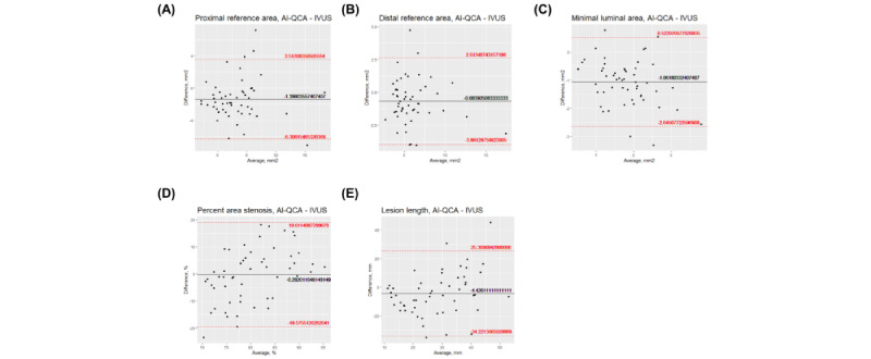
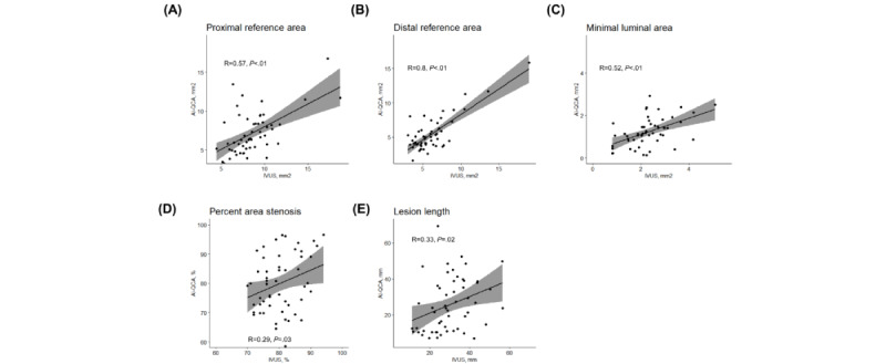
Results: A total of 54 significant lesions were analyzed in 47 patients. The proximal and distal reference areas, as well as the minimal luminal area, showed moderate to strong correlation between the 2 modalities (correlation coefficients of 0.57, 0.80, and 0.52, respectively; P<.001). The correlation was weaker for percent area stenosis and lesion length, although statistically significant (correlation coefficients of 0.29 and 0.33, respectively). AI-QCA tended to measure reference vessel areas smaller and lesion lengths shorter than IVUS did. Systemic proportional bias was not observed in Bland-Altman plots. The biggest cause of bias originated from the geographic mismatch of AI-QCA with IVUS. Discrepancies in the proximal or distal lesion margins were observed between the 2 modalities, which were more frequent at the distal margins. After the adjustment of proximal or distal margins, there was a stronger correlation of proximal and distal reference areas between AI-QCA and IVUS (correlation coefficients of 0.70 and 0.83, respectively).
Conclusions: AI-QCA showed a moderate to strong correlation compared with IVUS in analyzing coronary lesions with significant stenosis. The main discrepancy was in the perception of the distal margins by AI-QCA, and the correction of margins improved the correlation coefficients. We believe that this novel tool could provide confidence to treating physicians and help in making optimal clinical decisions.


 求助内容:
求助内容: 应助结果提醒方式:


