{"title":"An interpretable decision-support model for breast cancer diagnosis using histopathology images","authors":"Sruthi Krishna , S.S. Suganthi , Arnav Bhavsar , Jyotsna Yesodharan , Shivsubramani Krishnamoorthy","doi":"10.1016/j.jpi.2023.100319","DOIUrl":null,"url":null,"abstract":"<div><p>Microscopic examination of biopsy tissue slides is perceived as the gold-standard methodology for the confirmation of presence of cancer cells. Manual analysis of an overwhelming inflow of tissue slides is highly susceptible to misreading of tissue slides by pathologists. A computerized framework for histopathology image analysis is conceived as a diagnostic tool that greatly benefits pathologists, augmenting definitive diagnosis of cancer. Convolutional Neural Network (CNN) turned out to be the most adaptable and effective technique in the detection of abnormal pathologic histology. Despite their high sensitivity and predictive power, clinical translation is constrained by a lack of intelligible insights into the prediction. A computer-aided system that can offer a definitive diagnosis and interpretability is therefore highly desirable. Conventional visual explanatory techniques, Class Activation Mapping (CAM), combined with CNN models offers interpretable decision making. The major challenge in CAM is, it cannot be optimized to create the best visualization map. CAM also decreases the performance of the CNN models.</p><p>To address this challenge, we introduce a novel interpretable decision-support model using CNN with a trainable attention mechanism using response-based feed-forward visual explanation. We introduce a variant of DarkNet19 CNN model for the classification of histopathology images. In order to achieve visual interpretation as well as boost the performance of the DarkNet19 model, an attention branch is integrated with DarkNet19 network forming Attention Branch Network (ABN). The attention branch uses a convolution layer of DarkNet19 and Global Average Pooling (GAP) to model the context of the visual features and generate a heatmap to identify the region of interest. Finally, the perception branch is constituted using a fully connected layer to classify images.</p><p>We trained and validated our model using more than 7000 breast cancer biopsy slide images from an openly available dataset and achieved 98.7% accuracy in the binary classification of histopathology images. The observations substantiated the enhanced clinical interpretability of the DarkNet19 CNN model, supervened by the attention branch, besides delivering a 3%–4% performance boost of the baseline model. The cancer regions highlighted by the proposed model correlate well with the findings of an expert pathologist.</p><p>The coalesced approach of unifying attention branch with the CNN model capacitates pathologists with augmented diagnostic interpretability of histological images with no detriment to state-of-art performance. The model’s proficiency in pinpointing the region of interest is an added bonus that can lead to accurate clinical translation of deep learning models that underscore clinical decision support.</p></div>","PeriodicalId":37769,"journal":{"name":"Journal of Pathology Informatics","volume":"14 ","pages":"Article 100319"},"PeriodicalIF":0.0000,"publicationDate":"2023-01-01","publicationTypes":"Journal Article","fieldsOfStudy":null,"isOpenAccess":false,"openAccessPdf":"https://www.ncbi.nlm.nih.gov/pmc/articles/PMC10320615/pdf/","citationCount":"0","resultStr":null,"platform":"Semanticscholar","paperid":null,"PeriodicalName":"Journal of Pathology Informatics","FirstCategoryId":"1085","ListUrlMain":"https://www.sciencedirect.com/science/article/pii/S2153353923001335","RegionNum":0,"RegionCategory":null,"ArticlePicture":[],"TitleCN":null,"AbstractTextCN":null,"PMCID":null,"EPubDate":"","PubModel":"","JCR":"Q2","JCRName":"Medicine","Score":null,"Total":0}
引用次数: 0
Abstract
Microscopic examination of biopsy tissue slides is perceived as the gold-standard methodology for the confirmation of presence of cancer cells. Manual analysis of an overwhelming inflow of tissue slides is highly susceptible to misreading of tissue slides by pathologists. A computerized framework for histopathology image analysis is conceived as a diagnostic tool that greatly benefits pathologists, augmenting definitive diagnosis of cancer. Convolutional Neural Network (CNN) turned out to be the most adaptable and effective technique in the detection of abnormal pathologic histology. Despite their high sensitivity and predictive power, clinical translation is constrained by a lack of intelligible insights into the prediction. A computer-aided system that can offer a definitive diagnosis and interpretability is therefore highly desirable. Conventional visual explanatory techniques, Class Activation Mapping (CAM), combined with CNN models offers interpretable decision making. The major challenge in CAM is, it cannot be optimized to create the best visualization map. CAM also decreases the performance of the CNN models.
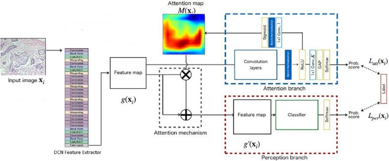
To address this challenge, we introduce a novel interpretable decision-support model using CNN with a trainable attention mechanism using response-based feed-forward visual explanation. We introduce a variant of DarkNet19 CNN model for the classification of histopathology images. In order to achieve visual interpretation as well as boost the performance of the DarkNet19 model, an attention branch is integrated with DarkNet19 network forming Attention Branch Network (ABN). The attention branch uses a convolution layer of DarkNet19 and Global Average Pooling (GAP) to model the context of the visual features and generate a heatmap to identify the region of interest. Finally, the perception branch is constituted using a fully connected layer to classify images.
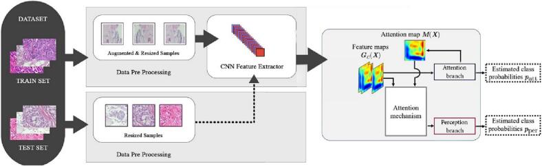
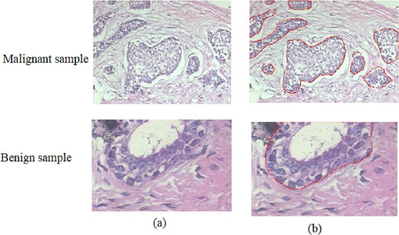
We trained and validated our model using more than 7000 breast cancer biopsy slide images from an openly available dataset and achieved 98.7% accuracy in the binary classification of histopathology images. The observations substantiated the enhanced clinical interpretability of the DarkNet19 CNN model, supervened by the attention branch, besides delivering a 3%–4% performance boost of the baseline model. The cancer regions highlighted by the proposed model correlate well with the findings of an expert pathologist.
The coalesced approach of unifying attention branch with the CNN model capacitates pathologists with augmented diagnostic interpretability of histological images with no detriment to state-of-art performance. The model’s proficiency in pinpointing the region of interest is an added bonus that can lead to accurate clinical translation of deep learning models that underscore clinical decision support.
期刊介绍:
The Journal of Pathology Informatics (JPI) is an open access peer-reviewed journal dedicated to the advancement of pathology informatics. This is the official journal of the Association for Pathology Informatics (API). The journal aims to publish broadly about pathology informatics and freely disseminate all articles worldwide. This journal is of interest to pathologists, informaticians, academics, researchers, health IT specialists, information officers, IT staff, vendors, and anyone with an interest in informatics. We encourage submissions from anyone with an interest in the field of pathology informatics. We publish all types of papers related to pathology informatics including original research articles, technical notes, reviews, viewpoints, commentaries, editorials, symposia, meeting abstracts, book reviews, and correspondence to the editors. All submissions are subject to rigorous peer review by the well-regarded editorial board and by expert referees in appropriate specialties.

 求助内容:
求助内容: 应助结果提醒方式:


