{"title":"Development of multifocal glioblastoma after radiotherapy for craniopharyngioma: illustrative case.","authors":"Edith Yuan, Michelle Lin, Elliot Min, Kristie Liu, Frank J Attenello","doi":"10.3171/CASE2377","DOIUrl":null,"url":null,"abstract":"<p><strong>Background: </strong>Radiation-induced glioblastoma (GBM) in patients previously treated for craniopharyngioma is a rare phenomenon. To the authors' knowledge, only seven cases have previously been documented in the literature.</p><p><strong>Observations: </strong>Herein, the authors report a case of a patient presenting with a new diagnosis of multifocal GBM 15 years after having received adjuvant radiotherapy for a craniopharyngioma. Magnetic resonance imaging revealed an extensive enhancing infiltrative lesion in the right frontal lobe as well as two satellite lesions in the contralateral frontal lobe. Histopathology on biopsy was consistent with GBM.</p><p><strong>Lessons: </strong>Even though this case is rare, it is nevertheless important to recognize GBM as a potential side effect of radiation. Long-term follow-up in postradiation craniopharyngioma patients is crucial for early detection.</p>","PeriodicalId":16554,"journal":{"name":"Journal of Neurosurgery: Case Lessons","volume":"5 26","pages":""},"PeriodicalIF":0.0000,"publicationDate":"2023-06-26","publicationTypes":"Journal Article","fieldsOfStudy":null,"isOpenAccess":false,"openAccessPdf":"https://ftp.ncbi.nlm.nih.gov/pub/pmc/oa_pdf/75/54/CASE2377.PMC10550543.pdf","citationCount":"0","resultStr":null,"platform":"Semanticscholar","paperid":null,"PeriodicalName":"Journal of Neurosurgery: Case Lessons","FirstCategoryId":"1085","ListUrlMain":"https://doi.org/10.3171/CASE2377","RegionNum":0,"RegionCategory":null,"ArticlePicture":[],"TitleCN":null,"AbstractTextCN":null,"PMCID":null,"EPubDate":"","PubModel":"","JCR":"","JCRName":"","Score":null,"Total":0}
引用次数: 0
Abstract
Background: Radiation-induced glioblastoma (GBM) in patients previously treated for craniopharyngioma is a rare phenomenon. To the authors' knowledge, only seven cases have previously been documented in the literature.
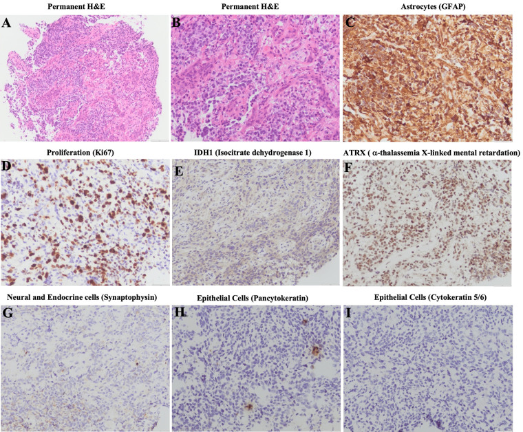
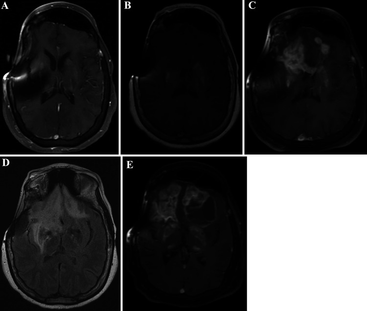
Observations: Herein, the authors report a case of a patient presenting with a new diagnosis of multifocal GBM 15 years after having received adjuvant radiotherapy for a craniopharyngioma. Magnetic resonance imaging revealed an extensive enhancing infiltrative lesion in the right frontal lobe as well as two satellite lesions in the contralateral frontal lobe. Histopathology on biopsy was consistent with GBM.
Lessons: Even though this case is rare, it is nevertheless important to recognize GBM as a potential side effect of radiation. Long-term follow-up in postradiation craniopharyngioma patients is crucial for early detection.

 求助内容:
求助内容: 应助结果提醒方式:


