Inamul Haque, Navanil Barua, Nabajyoti Borah, Sneha Gang, Ananya Barman, Shabnam A Ahmed
{"title":"Cervical Trident-Shaped Neurofibroma: A Rare Variant.","authors":"Inamul Haque, Navanil Barua, Nabajyoti Borah, Sneha Gang, Ananya Barman, Shabnam A Ahmed","doi":"10.1055/s-0043-1768579","DOIUrl":null,"url":null,"abstract":"<p><p>Spinal nerve root tumors can arise throughout the spine and at multiple levels, likely representing plexiform neurofibromas that grow from the nerve root into the intraspinal space either intradurally or epidurally and exit through the neural foramen, producing a dumbbell-shaped appearance. Although many cases of dumbbell-shaped extramedullary neurofibromas in the cervical spine have been reported, to the best of our knowledge, there are no reports of trident-shaped extramedullary neurofibromas. A 26-year-old woman presented with swelling over the right side of her neck. Diagnostic workup included magnetic resonance imaging (MRI) and contrast-enhanced computed tomography (CECT) of the neck, which revealed an intradural, extramedullary tumor mass at the right C2-C6 level with an extraspinal extension. Spinal cord compression or canal compromise is the most reliable indication for surgery. The solitary cervical neurofibroma was treated surgically in a single stage through laminoplasty and excision of the intradural tumor along with that of the neck component. This was performed without any complications. A single-stage double approach was adopted in this case. After total excision, the shape of the tumor was found to be more like a trident than a dumbbell. Hence, here we would like to suggest a new nomenclature for this neurofibroma, the trident neurofibroma.</p>","PeriodicalId":8521,"journal":{"name":"Asian Journal of Neurosurgery","volume":"18 2","pages":"357-365"},"PeriodicalIF":0.0000,"publicationDate":"2023-06-01","publicationTypes":"Journal Article","fieldsOfStudy":null,"isOpenAccess":false,"openAccessPdf":"https://ftp.ncbi.nlm.nih.gov/pub/pmc/oa_pdf/a0/f0/10-1055-s-0043-1768579.PMC10313435.pdf","citationCount":"0","resultStr":null,"platform":"Semanticscholar","paperid":null,"PeriodicalName":"Asian Journal of Neurosurgery","FirstCategoryId":"1085","ListUrlMain":"https://doi.org/10.1055/s-0043-1768579","RegionNum":0,"RegionCategory":null,"ArticlePicture":[],"TitleCN":null,"AbstractTextCN":null,"PMCID":null,"EPubDate":"","PubModel":"","JCR":"","JCRName":"","Score":null,"Total":0}
引用次数: 0
Abstract
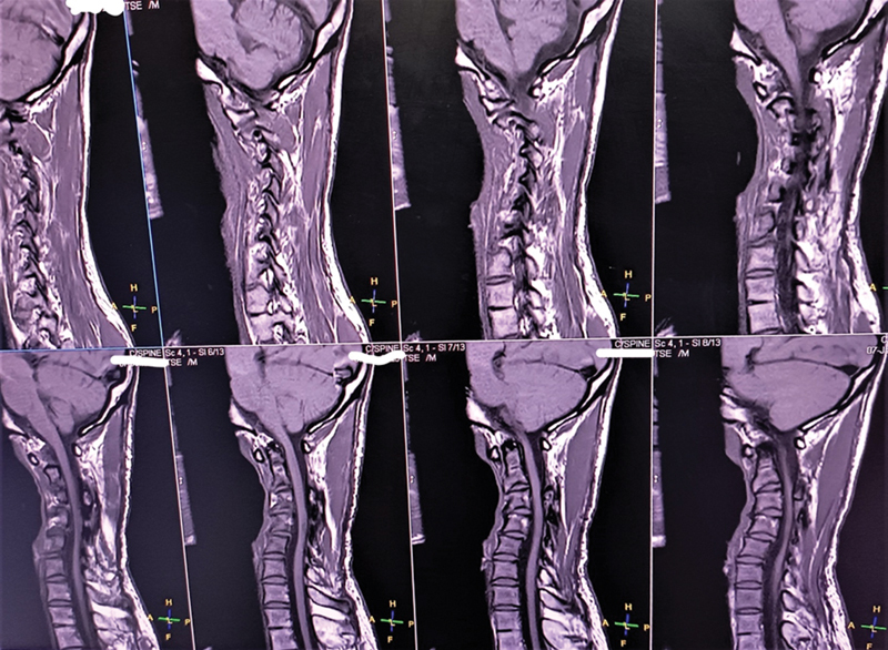
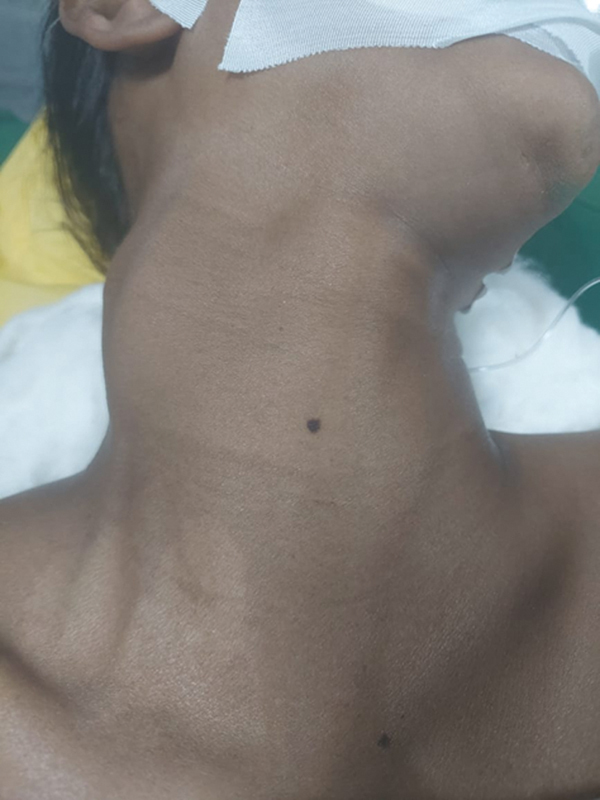
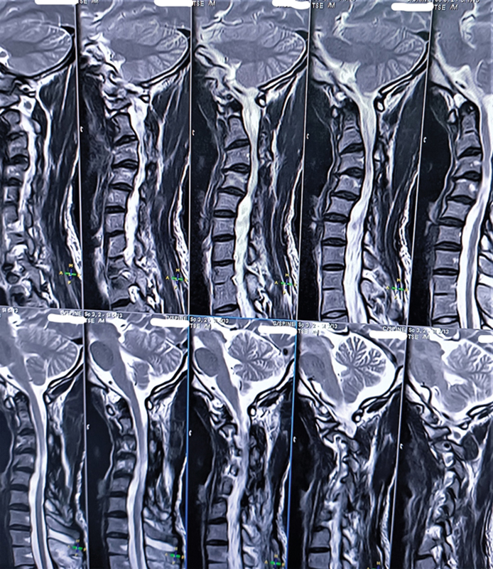
Spinal nerve root tumors can arise throughout the spine and at multiple levels, likely representing plexiform neurofibromas that grow from the nerve root into the intraspinal space either intradurally or epidurally and exit through the neural foramen, producing a dumbbell-shaped appearance. Although many cases of dumbbell-shaped extramedullary neurofibromas in the cervical spine have been reported, to the best of our knowledge, there are no reports of trident-shaped extramedullary neurofibromas. A 26-year-old woman presented with swelling over the right side of her neck. Diagnostic workup included magnetic resonance imaging (MRI) and contrast-enhanced computed tomography (CECT) of the neck, which revealed an intradural, extramedullary tumor mass at the right C2-C6 level with an extraspinal extension. Spinal cord compression or canal compromise is the most reliable indication for surgery. The solitary cervical neurofibroma was treated surgically in a single stage through laminoplasty and excision of the intradural tumor along with that of the neck component. This was performed without any complications. A single-stage double approach was adopted in this case. After total excision, the shape of the tumor was found to be more like a trident than a dumbbell. Hence, here we would like to suggest a new nomenclature for this neurofibroma, the trident neurofibroma.

 求助内容:
求助内容: 应助结果提醒方式:


