Lokeshwar S Bhenderu, Khaled M Taghlabi, Taimur Hassan, Jaime R Guerrero, Jesus G Cruz-Garza, Rachel L Goldstein, Shashank Sharma, Linda V Le, Tue A Dinh, Amir H Faraji
{"title":"Internal iliac artery aneurysm masquerading as a sciatic nerve schwannoma: illustrative case.","authors":"Lokeshwar S Bhenderu, Khaled M Taghlabi, Taimur Hassan, Jaime R Guerrero, Jesus G Cruz-Garza, Rachel L Goldstein, Shashank Sharma, Linda V Le, Tue A Dinh, Amir H Faraji","doi":"10.3171/CASE23175","DOIUrl":null,"url":null,"abstract":"<p><strong>Background: </strong>Schwannomas are common peripheral nerve sheath tumors. Imaging techniques such as magnetic resonance imaging (MRI) and computed tomography (CT) can help to distinguish schwannomas from other types of lesions. However, there have been several reported cases describing the misdiagnosis of aneurysms as schwannomas.</p><p><strong>Observations: </strong>A 70-year-old male with ongoing pain despite spinal fusion surgery underwent MRI. A lesion was noted along the left sciatic nerve, which was believed to be a sciatic nerve schwannoma. During the surgery for planned neurolysis and tumor resection, the lesion was noted to be pulsatile. Electromyography mapping and intraoperative ultrasound confirmed vascular pulsations and turbulent flow within the aneurysm, so the surgery was aborted. A formal CT angiogram revealed the lesion to be an internal iliac artery (IIA) branch aneurysm. The patient underwent coil embolization with complete obliteration of the aneurysm.</p><p><strong>Lessons: </strong>The authors report the first case of an IIA aneurysm misdiagnosed as a sciatic nerve schwannoma. Surgeons should be aware of this potential misdiagnosis and potentially use other imaging modalities to confirm the lesion before proceeding with surgery.</p>","PeriodicalId":16554,"journal":{"name":"Journal of Neurosurgery: Case Lessons","volume":"5 26","pages":""},"PeriodicalIF":0.0000,"publicationDate":"2023-06-26","publicationTypes":"Journal Article","fieldsOfStudy":null,"isOpenAccess":false,"openAccessPdf":"https://ftp.ncbi.nlm.nih.gov/pub/pmc/oa_pdf/22/50/CASE23175.PMC10550553.pdf","citationCount":"0","resultStr":null,"platform":"Semanticscholar","paperid":null,"PeriodicalName":"Journal of Neurosurgery: Case Lessons","FirstCategoryId":"1085","ListUrlMain":"https://doi.org/10.3171/CASE23175","RegionNum":0,"RegionCategory":null,"ArticlePicture":[],"TitleCN":null,"AbstractTextCN":null,"PMCID":null,"EPubDate":"","PubModel":"","JCR":"","JCRName":"","Score":null,"Total":0}
引用次数: 0
Abstract
Background: Schwannomas are common peripheral nerve sheath tumors. Imaging techniques such as magnetic resonance imaging (MRI) and computed tomography (CT) can help to distinguish schwannomas from other types of lesions. However, there have been several reported cases describing the misdiagnosis of aneurysms as schwannomas.
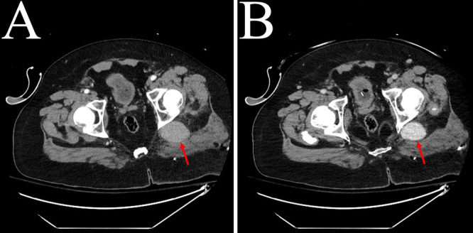
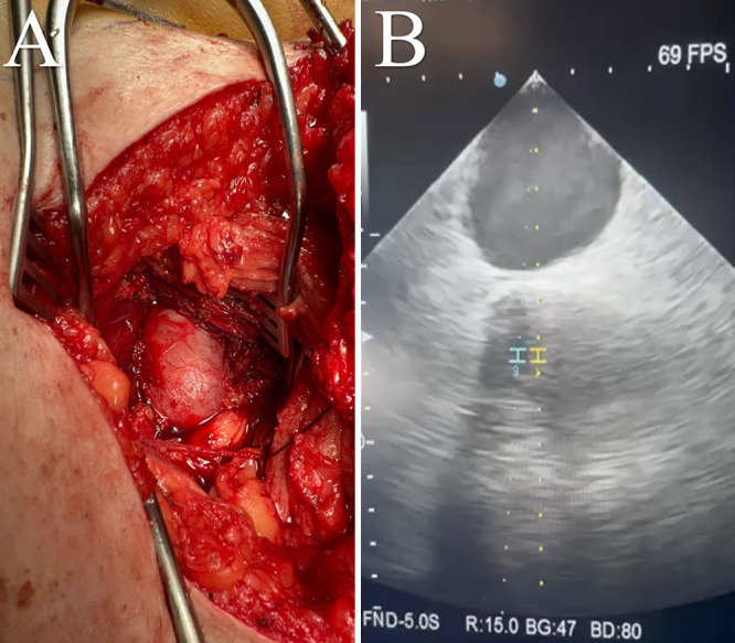
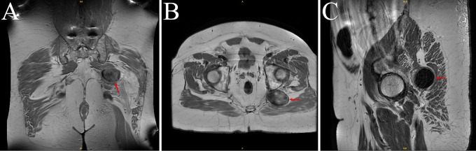
Observations: A 70-year-old male with ongoing pain despite spinal fusion surgery underwent MRI. A lesion was noted along the left sciatic nerve, which was believed to be a sciatic nerve schwannoma. During the surgery for planned neurolysis and tumor resection, the lesion was noted to be pulsatile. Electromyography mapping and intraoperative ultrasound confirmed vascular pulsations and turbulent flow within the aneurysm, so the surgery was aborted. A formal CT angiogram revealed the lesion to be an internal iliac artery (IIA) branch aneurysm. The patient underwent coil embolization with complete obliteration of the aneurysm.
Lessons: The authors report the first case of an IIA aneurysm misdiagnosed as a sciatic nerve schwannoma. Surgeons should be aware of this potential misdiagnosis and potentially use other imaging modalities to confirm the lesion before proceeding with surgery.

 求助内容:
求助内容: 应助结果提醒方式:


