A Rare Case of COVID-19-Associated Solitary Aspergillus Brain Abscess.
引用次数: 0
Abstract
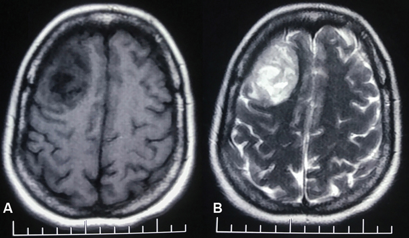
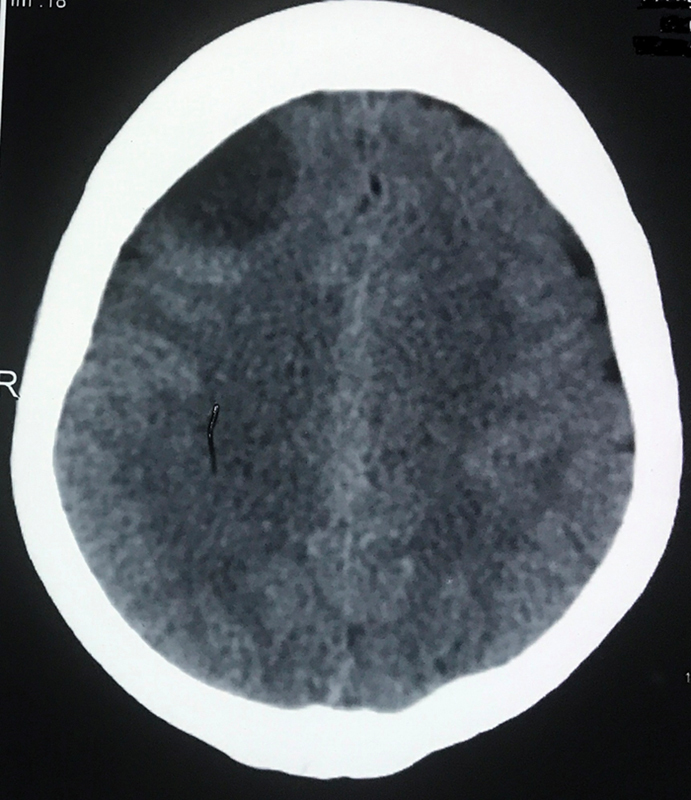
Abstract Surgically operated case of solitary Aspergillus brain abscess caused by Aspergillus fumigatus in coronavirus disease 2019 (COVID-19) patient is not reported. The authors report a case of 33-year-old diabetic female patient presented with generalized seizure followed by left hemiparesis. Patient was treated with steroids for COVID-19 pneumonia. Initial imaging revealed a right frontal lobe infarct that later confirmed as a case of frontal lobe abscess. Patient underwent craniotomy and thick yellow pus was drained. Abscess wall was excised. Postoperatively patient improved with Glasgow coma scale 15/15 and Medical Research Committee grade 5 power of all limbs. Microbiological examination of pus was done. The gram stain showed numerous pus cells with acute angle branching hyphae. Gomori methenamine silver (GMS) preparation showed filamentous black colored hyphae. Mycelial colonies appeared on chocolate agar after 48 hours of incubation. Cellophane tape mount from the plate showed conical shaped vesicle with conidia arising from the upper third of vesicle. Light green velvety colonies appeared on Sabouraud Dextrose Agar that later turned into smoky green. The isolate was identified as Aspergillus fumigatus . The hematoxylin and eosin stain of abscess wall section showed extensive areas of necrosis with few fungal hyphae. GMS stain of abscess wall showed fungal hyphae that are septate and showing acute angled branching which are consistent with Aspergillus species. Patient was treated with voriconazole. Imaging done after 8 months of surgery revealed no residue. Surgical excision of life-threatening solitary Aspergillus brain abscess along with antifungal medication voriconazole carries good result. The authors believe that decreased immunity in patient has contributed to the development of this rare disease. This is a rarest case of surgically operated solitary brain abscess caused by Aspergillus fumigatus in COVID-19 patient.


罕见的新冠肺炎相关性孤立曲霉性脑脓肿1例
新冠肺炎(COVID-19)患者由烟曲霉引起的孤立性曲霉性脑脓肿手术治疗一例未报道。作者报告了一例33岁的糖尿病女性患者,表现为全身性癫痫发作,随后出现左偏瘫。患者因COVID-19肺炎接受类固醇治疗。最初的影像显示为右额叶梗死,后来证实为额叶脓肿。病人接受开颅手术,排出厚厚的黄色脓液。切除脓肿壁。术后患者的格拉斯哥昏迷评分为15/15,医学研究委员会评分为5级。对脓液进行微生物学检查。革兰氏染色显示大量脓液细胞,有锐角分枝菌丝。Gomori甲基苯丙胺银(GMS)制备的菌丝呈丝状黑色。培养48小时后,在巧克力琼脂上出现菌丝菌落。玻璃纸贴片示圆锥形囊泡,囊泡上三分之一处有分生孢子。浅绿色丝绒菌落出现在Sabouraud葡萄糖琼脂上,后来变成烟绿色。该分离物经鉴定为烟曲霉。苏木精和伊红染色显示脓肿壁大面积坏死,真菌菌丝较少。脓肿壁GMS染色显示真菌菌丝分离,呈尖角分支,与曲霉属一致。患者给予伏立康唑治疗。手术8个月后影像学显示无残留。手术切除危及生命的孤立曲霉性脑脓肿,配合抗真菌药物伏立康唑治疗效果良好。作者认为,患者免疫力下降是导致这种罕见疾病发生的原因。这是COVID-19患者中最罕见的一例由烟曲霉引起的孤立性脑脓肿手术病例。
本文章由计算机程序翻译,如有差异,请以英文原文为准。
求助全文
约1分钟内获得全文
求助全文

 求助内容:
求助内容: 应助结果提醒方式:


