{"title":"Coincident Acute Macular Neuroretinopathy and Paracentral Acute Middle Maculopathy in COVID-19.","authors":"Aslıhan Yılmaz Çebi, Oğuzhan Kılıçarslan, Didar Uçar","doi":"10.4274/tjo.galenos.2022.55156","DOIUrl":null,"url":null,"abstract":"<p><p>An ophthalmology consultation was requested for a 29-year-old woman complaining of visual field defects. The patient had presented to the emergency department with cough and high fever one day earlier. Chest computed tomography demonstrated pneumonia and two severe acute respiratory syndrome coronavirus 2 polymerase chain reaction tests were positive. The patient had undergone renal transplantation 11 years ago due to glomerulonephritis. Best-corrected visual acuity (BCVA) was 20/40 in the right eye and 20/30 in the left eye. Fluorescein angiography showed macular hypoperfusion, and optical coherence tomography (OCT) showed hyperreflectivity in the inner nuclear, outer plexiform, and outer nuclear layers, as well as discontinuity of the ellipsoid zone. Perimetry confirmed bilateral central scotoma. Levels of D-dimer and fibrinogen were 0.86 g/mL and 435.6 g/mL, respectively. The patient was diagnosed as having concurrent acute macular neuroretinopathy and paracentral acute middle maculopathy and was given low-molecular-weight heparin treatment for one month. Her BCVA improved to 20/20 in both eyes, and regression was observed in the retinal findings, hyperreflectivity and ellipsoid zone disruption on OCT, and scotoma in perimetry. Inflammation, thrombosis, and glial involvement may play a role in the pathogenesis of retinal microvascular impairment in COVID-19.</p>","PeriodicalId":23373,"journal":{"name":"Turkish Journal of Ophthalmology","volume":null,"pages":null},"PeriodicalIF":0.0000,"publicationDate":"2023-04-20","publicationTypes":"Journal Article","fieldsOfStudy":null,"isOpenAccess":false,"openAccessPdf":"https://ftp.ncbi.nlm.nih.gov/pub/pmc/oa_pdf/b7/99/TJO-53-120.PMC10127542.pdf","citationCount":"1","resultStr":null,"platform":"Semanticscholar","paperid":null,"PeriodicalName":"Turkish Journal of Ophthalmology","FirstCategoryId":"1085","ListUrlMain":"https://doi.org/10.4274/tjo.galenos.2022.55156","RegionNum":0,"RegionCategory":null,"ArticlePicture":[],"TitleCN":null,"AbstractTextCN":null,"PMCID":null,"EPubDate":"","PubModel":"","JCR":"Q3","JCRName":"Medicine","Score":null,"Total":0}
引用次数: 1
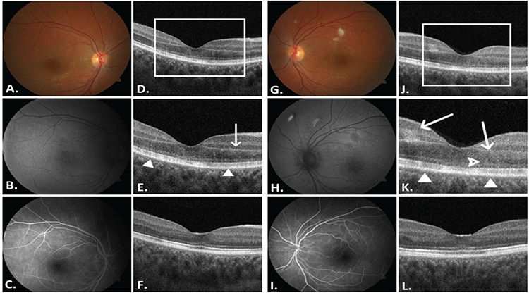
Abstract
An ophthalmology consultation was requested for a 29-year-old woman complaining of visual field defects. The patient had presented to the emergency department with cough and high fever one day earlier. Chest computed tomography demonstrated pneumonia and two severe acute respiratory syndrome coronavirus 2 polymerase chain reaction tests were positive. The patient had undergone renal transplantation 11 years ago due to glomerulonephritis. Best-corrected visual acuity (BCVA) was 20/40 in the right eye and 20/30 in the left eye. Fluorescein angiography showed macular hypoperfusion, and optical coherence tomography (OCT) showed hyperreflectivity in the inner nuclear, outer plexiform, and outer nuclear layers, as well as discontinuity of the ellipsoid zone. Perimetry confirmed bilateral central scotoma. Levels of D-dimer and fibrinogen were 0.86 g/mL and 435.6 g/mL, respectively. The patient was diagnosed as having concurrent acute macular neuroretinopathy and paracentral acute middle maculopathy and was given low-molecular-weight heparin treatment for one month. Her BCVA improved to 20/20 in both eyes, and regression was observed in the retinal findings, hyperreflectivity and ellipsoid zone disruption on OCT, and scotoma in perimetry. Inflammation, thrombosis, and glial involvement may play a role in the pathogenesis of retinal microvascular impairment in COVID-19.
期刊介绍:
The Turkish Journal of Ophthalmology (TJO) is the only scientific periodical publication of the Turkish Ophthalmological Association and has been published since January 1929. In its early years, the journal was published in Turkish and French. Although there were temporary interruptions in the publication of the journal due to various challenges, the Turkish Journal of Ophthalmology has been published continually from 1971 to the present. The target audience includes specialists and physicians in training in ophthalmology in all relevant disciplines.

 求助内容:
求助内容: 应助结果提醒方式:


