Gökçen Deniz Gülpınar İkiz, Ece Özdemir Zeydanlı, Şengül Özdek
{"title":"Flap-Related Complications Following Temporal Inverted Internal Limiting Membrane Flap for Macular Hole Repair.","authors":"Gökçen Deniz Gülpınar İkiz, Ece Özdemir Zeydanlı, Şengül Özdek","doi":"10.4274/tjo.galenos.2022.04828","DOIUrl":null,"url":null,"abstract":"<p><p>Here we report three cases of flap-related complications following temporal inverted internal limiting membrane (ILM) flap technique for the repair of macular holes (MH). The first case showed a flap closure pattern in which the MH completely closed at 2 months spontaneously. The second case showed early anatomical and functional improvement provided by an immediate closure of the MH but developed flap contracture and nasally located epiretinal membrane (ERM) at postoperative 18 months. There was no functional deterioration, thus no further intervention was required. In the third case, early postoperative flap dislocation was observed and an additional surgery to reposition the flap was needed. The flap closure pattern observed with inverted ILM flap techniques may represent the ongoing healing process of large MHs and may be related to delayed spontaneous anatomical closure. ILM flap contracture and ERM formation may be a harmless long-term complication. Dislocation of the ILM flap is an unexpected early postoperative complication that may necessitate a second surgery for flap repositioning.</p>","PeriodicalId":23373,"journal":{"name":"Turkish Journal of Ophthalmology","volume":null,"pages":null},"PeriodicalIF":0.0000,"publicationDate":"2023-04-20","publicationTypes":"Journal Article","fieldsOfStudy":null,"isOpenAccess":false,"openAccessPdf":"https://ftp.ncbi.nlm.nih.gov/pub/pmc/oa_pdf/de/e5/TJO-53-130.PMC10127537.pdf","citationCount":"0","resultStr":null,"platform":"Semanticscholar","paperid":null,"PeriodicalName":"Turkish Journal of Ophthalmology","FirstCategoryId":"1085","ListUrlMain":"https://doi.org/10.4274/tjo.galenos.2022.04828","RegionNum":0,"RegionCategory":null,"ArticlePicture":[],"TitleCN":null,"AbstractTextCN":null,"PMCID":null,"EPubDate":"","PubModel":"","JCR":"Q3","JCRName":"Medicine","Score":null,"Total":0}
引用次数: 0
Abstract
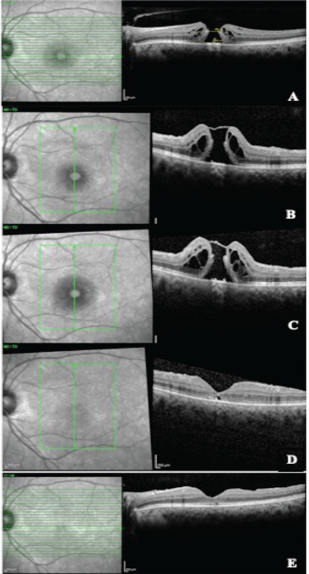
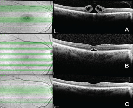
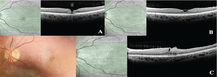
Here we report three cases of flap-related complications following temporal inverted internal limiting membrane (ILM) flap technique for the repair of macular holes (MH). The first case showed a flap closure pattern in which the MH completely closed at 2 months spontaneously. The second case showed early anatomical and functional improvement provided by an immediate closure of the MH but developed flap contracture and nasally located epiretinal membrane (ERM) at postoperative 18 months. There was no functional deterioration, thus no further intervention was required. In the third case, early postoperative flap dislocation was observed and an additional surgery to reposition the flap was needed. The flap closure pattern observed with inverted ILM flap techniques may represent the ongoing healing process of large MHs and may be related to delayed spontaneous anatomical closure. ILM flap contracture and ERM formation may be a harmless long-term complication. Dislocation of the ILM flap is an unexpected early postoperative complication that may necessitate a second surgery for flap repositioning.
期刊介绍:
The Turkish Journal of Ophthalmology (TJO) is the only scientific periodical publication of the Turkish Ophthalmological Association and has been published since January 1929. In its early years, the journal was published in Turkish and French. Although there were temporary interruptions in the publication of the journal due to various challenges, the Turkish Journal of Ophthalmology has been published continually from 1971 to the present. The target audience includes specialists and physicians in training in ophthalmology in all relevant disciplines.

 求助内容:
求助内容: 应助结果提醒方式:


