Mansour Mathkour, Cassidy D Werner, Robert F Dallapiazza, Marios Loukas, Joe Iwanaga, Aaron S Dumont, R Shane Tubbs
{"title":"Endoscopically-Assisted Percutaneous Trigeminal Rhizotomy for Trigeminal Neuralgia: A Cadaveric Feasibility Study.","authors":"Mansour Mathkour, Cassidy D Werner, Robert F Dallapiazza, Marios Loukas, Joe Iwanaga, Aaron S Dumont, R Shane Tubbs","doi":"10.1055/s-0043-1761230","DOIUrl":null,"url":null,"abstract":"<p><p><b>Objective</b> Trigeminal neuralgia (TN) is a debilitating syndrome characterized by paroxysmal facial pain in one or more divisions of the trigeminal nerve. The etiology and treatment paradigms are still controversial. The endoscopically-assisted procedure has not yet been described in percutaneous procedures for TN. The aim of this study was to assess the utility and feasibility of endoscopic-assisted percutaneous approaches for trigeminal rhizotomy in TN. <b>Methods</b> This study comprised eight cadaveric sides heads that underwent an endoscopically assisted percutaneous approach using Hakanson's anterior puncture method for targeting the foramen ovale. <b>Results</b> V3 exiting the foramen ovale was easily visualized with the endoscope on all sides. While approaching the foramen ovale, distal branches of V3 such as the lingual and inferior alveolar nerves were first identified as they traveled between the medial and lateral pterygoid muscles. These branches were then traced proximally to the V3 trunk deep to the lateral pterygoid. Large arteries and veins were easily visualized and avoided in the trajectory to the foramen ovale. No gross injury to any neurovascular structure along the course of the needle insertion was identified. <b>Conclusion</b> We found that endoscopic-assisted percutaneous approach to the foramen ovale is feasible and allows for accurate canalization and anatomical identification of the precise location for rhizotomy under direct visualization. Such a procedure, after it is confirmed in patients, could offer a new technique for reducing unsuccessful canalization and could improve outcomes.</p>","PeriodicalId":8521,"journal":{"name":"Asian Journal of Neurosurgery","volume":"18 1","pages":"40-44"},"PeriodicalIF":0.0000,"publicationDate":"2023-03-01","publicationTypes":"Journal Article","fieldsOfStudy":null,"isOpenAccess":false,"openAccessPdf":"https://ftp.ncbi.nlm.nih.gov/pub/pmc/oa_pdf/61/e2/10-1055-s-0043-1761230.PMC10089747.pdf","citationCount":"0","resultStr":null,"platform":"Semanticscholar","paperid":null,"PeriodicalName":"Asian Journal of Neurosurgery","FirstCategoryId":"1085","ListUrlMain":"https://doi.org/10.1055/s-0043-1761230","RegionNum":0,"RegionCategory":null,"ArticlePicture":[],"TitleCN":null,"AbstractTextCN":null,"PMCID":null,"EPubDate":"","PubModel":"","JCR":"","JCRName":"","Score":null,"Total":0}
引用次数: 0
Abstract
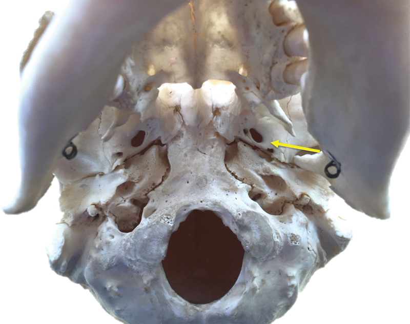
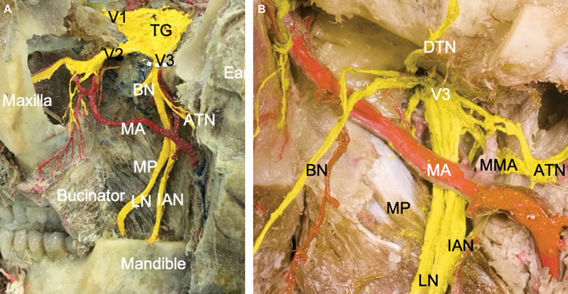
Objective Trigeminal neuralgia (TN) is a debilitating syndrome characterized by paroxysmal facial pain in one or more divisions of the trigeminal nerve. The etiology and treatment paradigms are still controversial. The endoscopically-assisted procedure has not yet been described in percutaneous procedures for TN. The aim of this study was to assess the utility and feasibility of endoscopic-assisted percutaneous approaches for trigeminal rhizotomy in TN. Methods This study comprised eight cadaveric sides heads that underwent an endoscopically assisted percutaneous approach using Hakanson's anterior puncture method for targeting the foramen ovale. Results V3 exiting the foramen ovale was easily visualized with the endoscope on all sides. While approaching the foramen ovale, distal branches of V3 such as the lingual and inferior alveolar nerves were first identified as they traveled between the medial and lateral pterygoid muscles. These branches were then traced proximally to the V3 trunk deep to the lateral pterygoid. Large arteries and veins were easily visualized and avoided in the trajectory to the foramen ovale. No gross injury to any neurovascular structure along the course of the needle insertion was identified. Conclusion We found that endoscopic-assisted percutaneous approach to the foramen ovale is feasible and allows for accurate canalization and anatomical identification of the precise location for rhizotomy under direct visualization. Such a procedure, after it is confirmed in patients, could offer a new technique for reducing unsuccessful canalization and could improve outcomes.


 求助内容:
求助内容: 应助结果提醒方式:


