{"title":"Gingko Leaf Sign: A Classical Imaging Finding in Spinal Meningiomas.","authors":"Prasad Krishnan","doi":"10.1055/s-0043-1760853","DOIUrl":null,"url":null,"abstract":"<p><p>The common imaging features surgeons use to distinguish spinal meningiomas from spinal nerve sheath tumors on magnetic resonance (MR) scans include the presence of the \"dural tail sign\" on contrast-enhanced MR images, hypointensity of the lesion on T2 sequences, presence of calcifications, lack of extraspinal dumbbell extension, and the lack of cystic changes in the lesion. We highlight the rarely described finding-the \"Gingko-Leaf\" sign that is caused by the stretched denticulate ligament as it extends laterally, through the tumor, from the compressed spinal cord.</p>","PeriodicalId":8521,"journal":{"name":"Asian Journal of Neurosurgery","volume":"18 1","pages":"228-229"},"PeriodicalIF":0.0000,"publicationDate":"2023-03-01","publicationTypes":"Journal Article","fieldsOfStudy":null,"isOpenAccess":false,"openAccessPdf":"https://ftp.ncbi.nlm.nih.gov/pub/pmc/oa_pdf/dc/a2/10-1055-s-0043-1760853.PMC10089726.pdf","citationCount":"0","resultStr":null,"platform":"Semanticscholar","paperid":null,"PeriodicalName":"Asian Journal of Neurosurgery","FirstCategoryId":"1085","ListUrlMain":"https://doi.org/10.1055/s-0043-1760853","RegionNum":0,"RegionCategory":null,"ArticlePicture":[],"TitleCN":null,"AbstractTextCN":null,"PMCID":null,"EPubDate":"","PubModel":"","JCR":"","JCRName":"","Score":null,"Total":0}
引用次数: 0
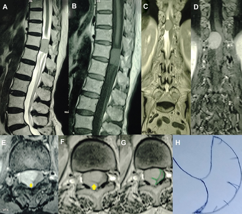
Abstract
The common imaging features surgeons use to distinguish spinal meningiomas from spinal nerve sheath tumors on magnetic resonance (MR) scans include the presence of the "dural tail sign" on contrast-enhanced MR images, hypointensity of the lesion on T2 sequences, presence of calcifications, lack of extraspinal dumbbell extension, and the lack of cystic changes in the lesion. We highlight the rarely described finding-the "Gingko-Leaf" sign that is caused by the stretched denticulate ligament as it extends laterally, through the tumor, from the compressed spinal cord.

 求助内容:
求助内容: 应助结果提醒方式:


