Knockdown of Yap attenuates TAA-induced hepatic fibrosis by interaction with hedgehog signals.

IF 3.6 3区 生物学 Q3 CELL BIOLOGY
Ye Zhao, Huiling Wang, Tianhua He, Bo Ma, Guoguang Chen, Chimeng Tzeng
{"title":"Knockdown of Yap attenuates TAA-induced hepatic fibrosis by interaction with hedgehog signals.","authors":"Ye Zhao, Huiling Wang, Tianhua He, Bo Ma, Guoguang Chen, Chimeng Tzeng","doi":"10.1007/s12079-023-00775-6","DOIUrl":null,"url":null,"abstract":"<p><p>Liver fibrosis is an aberrant wound healing response to tissue injury characterized by excessive extracellular matrix deposition and loss of normal liver architecture. Hepatic stellate cells (HSCs) activation is regards to be the major process in liver fibrogenesis which is dynamic and reversible. Both Hippo signaling core factor Yap and Hedgehog (Hh) signaling promote HSCs transdifferentiation thereby regulating the repair process of liver injury. However, the molecular function of YAP and the regulation between Yap and Hh during fibrogenesis remain uncertain. In this study, the essential roles of Yap in liver fibrosis were investigated. Yap was detected to be increased in liver fibrotic tissue by the thioacetamide (TAA)-induced zebrafish embryonic and adult models. Inhibition of Yap by both embryonic morpholino interference and adult's inhibitor treatment was proved to alleviate TAA-induced liver lesions by and histology and gene expression examination. Transcriptomic analysis and gene expression detection showed that Yap and Hh signaling pathway have a cross talking upon TAA-induced liver fibrosis. In addition, TAA induction promoted the nuclear colocalization of YAP and Hh signaling factor GLI2α. This study demonstrates that Yap and Hh play synergistic protective roles in liver fibrotic response and provides new theoretical insight concerning the mechanisms of fibrosis progression.</p>","PeriodicalId":15226,"journal":{"name":"Journal of Cell Communication and Signaling","volume":" ","pages":"1335-1354"},"PeriodicalIF":3.6000,"publicationDate":"2023-12-01","publicationTypes":"Journal Article","fieldsOfStudy":null,"isOpenAccess":false,"openAccessPdf":"https://www.ncbi.nlm.nih.gov/pmc/articles/PMC10713942/pdf/","citationCount":"1","resultStr":null,"platform":"Semanticscholar","paperid":null,"PeriodicalName":"Journal of Cell Communication and Signaling","FirstCategoryId":"99","ListUrlMain":"https://doi.org/10.1007/s12079-023-00775-6","RegionNum":3,"RegionCategory":"生物学","ArticlePicture":[],"TitleCN":null,"AbstractTextCN":null,"PMCID":null,"EPubDate":"2023/6/20 0:00:00","PubModel":"Epub","JCR":"Q3","JCRName":"CELL BIOLOGY","Score":null,"Total":0}
引用次数: 1

Abstract

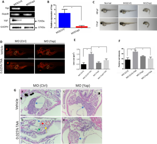
Liver fibrosis is an aberrant wound healing response to tissue injury characterized by excessive extracellular matrix deposition and loss of normal liver architecture. Hepatic stellate cells (HSCs) activation is regards to be the major process in liver fibrogenesis which is dynamic and reversible. Both Hippo signaling core factor Yap and Hedgehog (Hh) signaling promote HSCs transdifferentiation thereby regulating the repair process of liver injury. However, the molecular function of YAP and the regulation between Yap and Hh during fibrogenesis remain uncertain. In this study, the essential roles of Yap in liver fibrosis were investigated. Yap was detected to be increased in liver fibrotic tissue by the thioacetamide (TAA)-induced zebrafish embryonic and adult models. Inhibition of Yap by both embryonic morpholino interference and adult's inhibitor treatment was proved to alleviate TAA-induced liver lesions by and histology and gene expression examination. Transcriptomic analysis and gene expression detection showed that Yap and Hh signaling pathway have a cross talking upon TAA-induced liver fibrosis. In addition, TAA induction promoted the nuclear colocalization of YAP and Hh signaling factor GLI2α. This study demonstrates that Yap and Hh play synergistic protective roles in liver fibrotic response and provides new theoretical insight concerning the mechanisms of fibrosis progression.
Abstract Image

通过与刺猬信号相互作用,敲除 Yap 可减轻 TAA 诱导的肝纤维化。
肝纤维化是对组织损伤的一种异常伤口愈合反应,其特点是细胞外基质过度沉积和正常肝脏结构丧失。肝星状细胞(HSCs)活化被认为是肝纤维化的主要过程,而肝纤维化是动态和可逆的。Hippo信号转导核心因子Yap和Hedgehog(Hh)信号转导都能促进造血干细胞的转分化,从而调节肝损伤的修复过程。然而,YAP的分子功能以及Yap和Hh在纤维化过程中的调控作用仍不确定。本研究探讨了Yap在肝纤维化中的重要作用。硫代乙酰胺(TAA)诱导的斑马鱼胚胎和成鱼模型检测到肝纤维化组织中的Yap增加。通过组织学和基因表达检测,证明通过胚胎吗啉干扰和成鱼抑制剂处理抑制Yap可减轻TAA诱导的肝脏病变。转录组分析和基因表达检测表明,Yap和Hh信号通路在TAA诱导的肝纤维化过程中存在交叉对话。此外,TAA诱导促进了YAP和Hh信号因子GLI2α的核共定位。该研究表明,Yap和Hh在肝纤维化反应中发挥协同保护作用,并为肝纤维化进展机制提供了新的理论依据。
本文章由计算机程序翻译,如有差异,请以英文原文为准。
求助全文
约1分钟内获得全文 求助全文
来源期刊
CiteScore
6.40
自引率
4.90%
发文量
40
期刊介绍: The Journal of Cell Communication and Signaling provides a forum for fundamental and translational research. In particular, it publishes papers discussing intercellular and intracellular signaling pathways that are particularly important to understand how cells interact with each other and with the surrounding environment, and how cellular behavior contributes to pathological states. JCCS encourages the submission of research manuscripts, timely reviews and short commentaries discussing recent publications, key developments and controversies. Research manuscripts can be published under two different sections : In the Pathology and Translational Research Section (Section Editor Andrew Leask) , manuscripts report original research dealing with celllular aspects of normal and pathological signaling and communication, with a particular interest in translational research. In the Molecular Signaling Section (Section Editor Satoshi Kubota) manuscripts report original signaling research performed at molecular levels with a particular interest in the functions of intracellular and membrane components involved in cell signaling.
×
引用
GB/T 7714-2015
复制
MLA
复制
APA
复制
导出至
BibTeX EndNote RefMan NoteFirst NoteExpress
×
提示
您的信息不完整,为了账户安全,请先补充。
现在去补充
×
提示
您因"违规操作"
具体请查看互助需知
我知道了
×
提示
确定
请完成安全验证×
copy
已复制链接
快去分享给好友吧!
我知道了
右上角分享
点击右上角分享
0
联系我们:info@booksci.cn Book学术提供免费学术资源搜索服务,方便国内外学者检索中英文文献。致力于提供最便捷和优质的服务体验。 Copyright © 2023 布克学术 All rights reserved.
京ICP备2023020795号-1
ghs 京公网安备 11010802042870号
Book学术文献互助
Book学术文献互助群
群 号:481959085
Book学术官方微信