Kristina F Terrani, Anthony M Avellino, M Michael Bercu
{"title":"Enlarging traumatic superficial temporal artery pseudoaneurysm from a lacrosse ball injury: illustrative case.","authors":"Kristina F Terrani, Anthony M Avellino, M Michael Bercu","doi":"10.3171/CASE2339","DOIUrl":null,"url":null,"abstract":"<p><strong>Background: </strong>The development of a mobile, growing, pulsatile mass after blunt head trauma to the forehead area, resulting in a superficial temporal artery pseudoaneurysm, is a very rare outcome. Most pseudoaneurysms are diagnosed with ultrasound, computed tomography (CT), and/or magnetic resonance imaging and treated via resection or, occasionally, embolization.</p><p><strong>Observations: </strong>The authors describe a case of a young male lacrosse player who presented with a bulging, partially pulsatile mass in the right forehead region 2 months after trauma from a high-velocity ball striking his head while helmeted. The authors reviewed 12 patients in the literature and describe each patient's epidemiological features, nature of the trauma, and onset of the lesion after the trauma, as well as the diagnostic methods and treatments for each case.</p><p><strong>Lessons: </strong>Overall, CT and ultrasound appear to be the easiest and most used methods of diagnosis, and resection under general anesthesia is the most common treatment method.</p>","PeriodicalId":16554,"journal":{"name":"Journal of Neurosurgery: Case Lessons","volume":"5 24","pages":""},"PeriodicalIF":0.0000,"publicationDate":"2023-06-12","publicationTypes":"Journal Article","fieldsOfStudy":null,"isOpenAccess":false,"openAccessPdf":"https://ftp.ncbi.nlm.nih.gov/pub/pmc/oa_pdf/82/4f/CASE2339.PMC10550658.pdf","citationCount":"0","resultStr":null,"platform":"Semanticscholar","paperid":null,"PeriodicalName":"Journal of Neurosurgery: Case Lessons","FirstCategoryId":"1085","ListUrlMain":"https://doi.org/10.3171/CASE2339","RegionNum":0,"RegionCategory":null,"ArticlePicture":[],"TitleCN":null,"AbstractTextCN":null,"PMCID":null,"EPubDate":"","PubModel":"","JCR":"","JCRName":"","Score":null,"Total":0}
引用次数: 0
Abstract
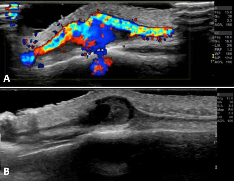
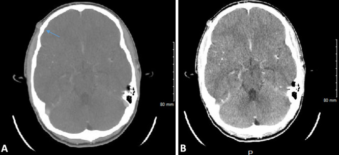
Background: The development of a mobile, growing, pulsatile mass after blunt head trauma to the forehead area, resulting in a superficial temporal artery pseudoaneurysm, is a very rare outcome. Most pseudoaneurysms are diagnosed with ultrasound, computed tomography (CT), and/or magnetic resonance imaging and treated via resection or, occasionally, embolization.
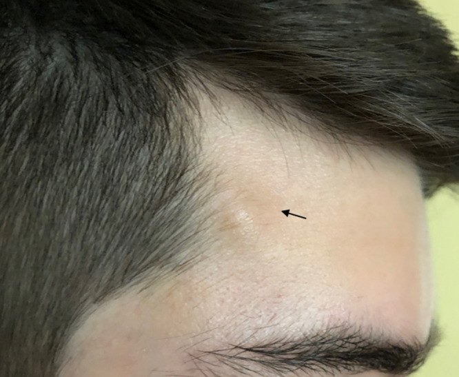
Observations: The authors describe a case of a young male lacrosse player who presented with a bulging, partially pulsatile mass in the right forehead region 2 months after trauma from a high-velocity ball striking his head while helmeted. The authors reviewed 12 patients in the literature and describe each patient's epidemiological features, nature of the trauma, and onset of the lesion after the trauma, as well as the diagnostic methods and treatments for each case.
Lessons: Overall, CT and ultrasound appear to be the easiest and most used methods of diagnosis, and resection under general anesthesia is the most common treatment method.

 求助内容:
求助内容: 应助结果提醒方式:


