Hye Jin Park, Ji Young Lee, Jin-Ju Yang, Hee-Jin Kim, Young Seo Kim, Ji Young Kim, Yun Young Choi
{"title":"Prediction of Amyloid β-Positivity with both MRI Parameters and Cognitive Function Using Machine Learning.","authors":"Hye Jin Park, Ji Young Lee, Jin-Ju Yang, Hee-Jin Kim, Young Seo Kim, Ji Young Kim, Yun Young Choi","doi":"10.3348/jksr.2022.0084","DOIUrl":null,"url":null,"abstract":"<p><strong>Purpose: </strong>To investigate the MRI markers for the prediction of amyloid β (Aβ)-positivity in mild cognitive impairment (MCI) and Alzheimer's disease (AD), and to evaluate the differences in MRI markers between Aβ-positive (Aβ [+]) and -negative groups using the machine learning (ML) method.</p><p><strong>Materials and methods: </strong>This study included 139 patients with MCI and AD who underwent amyloid PET-CT and brain MRI. Patients were divided into Aβ (+) (<i>n</i> = 84) and Aβ-negative (<i>n</i> = 55) groups. Visual analysis was performed with the Fazekas scale of white matter hyperintensity (WMH) and cerebral microbleeds (CMB) scores. The WMH volume and regional brain volume were quantitatively measured. The multivariable logistic regression and ML using support vector machine, and logistic regression were used to identify the best MRI predictors of Aβ-positivity.</p><p><strong>Results: </strong>The Fazekas scale of WMH (<i>p</i> = 0.02) and CMB scores (<i>p</i> = 0.04) were higher in Aβ (+). The volumes of hippocampus, entorhinal cortex, and precuneus were smaller in Aβ (+) (<i>p</i> < 0.05). The third ventricle volume was larger in Aβ (+) (<i>p</i> = 0.002). The logistic regression of ML showed a good accuracy (81.1%) with mini-mental state examination (MMSE) and regional brain volumes.</p><p><strong>Conclusion: </strong>The application of ML using the MMSE, third ventricle, and hippocampal volume is helpful in predicting Aβ-positivity with a good accuracy.</p>","PeriodicalId":17455,"journal":{"name":"Journal of the Korean Society of Radiology","volume":"84 3","pages":"638-652"},"PeriodicalIF":0.0000,"publicationDate":"2023-05-01","publicationTypes":"Journal Article","fieldsOfStudy":null,"isOpenAccess":false,"openAccessPdf":"https://ftp.ncbi.nlm.nih.gov/pub/pmc/oa_pdf/3e/34/jksr-84-638.PMC10265247.pdf","citationCount":"0","resultStr":null,"platform":"Semanticscholar","paperid":null,"PeriodicalName":"Journal of the Korean Society of Radiology","FirstCategoryId":"1085","ListUrlMain":"https://doi.org/10.3348/jksr.2022.0084","RegionNum":0,"RegionCategory":null,"ArticlePicture":[],"TitleCN":null,"AbstractTextCN":null,"PMCID":null,"EPubDate":"","PubModel":"","JCR":"Q4","JCRName":"Medicine","Score":null,"Total":0}
引用次数: 0
Abstract
Purpose: To investigate the MRI markers for the prediction of amyloid β (Aβ)-positivity in mild cognitive impairment (MCI) and Alzheimer's disease (AD), and to evaluate the differences in MRI markers between Aβ-positive (Aβ [+]) and -negative groups using the machine learning (ML) method.
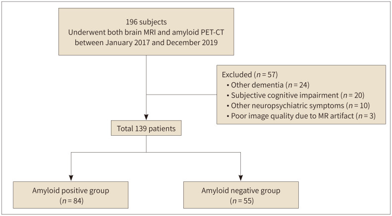
Materials and methods: This study included 139 patients with MCI and AD who underwent amyloid PET-CT and brain MRI. Patients were divided into Aβ (+) (n = 84) and Aβ-negative (n = 55) groups. Visual analysis was performed with the Fazekas scale of white matter hyperintensity (WMH) and cerebral microbleeds (CMB) scores. The WMH volume and regional brain volume were quantitatively measured. The multivariable logistic regression and ML using support vector machine, and logistic regression were used to identify the best MRI predictors of Aβ-positivity.
Results: The Fazekas scale of WMH (p = 0.02) and CMB scores (p = 0.04) were higher in Aβ (+). The volumes of hippocampus, entorhinal cortex, and precuneus were smaller in Aβ (+) (p < 0.05). The third ventricle volume was larger in Aβ (+) (p = 0.002). The logistic regression of ML showed a good accuracy (81.1%) with mini-mental state examination (MMSE) and regional brain volumes.
Conclusion: The application of ML using the MMSE, third ventricle, and hippocampal volume is helpful in predicting Aβ-positivity with a good accuracy.

 求助内容:
求助内容: 应助结果提醒方式:


