Hyun Jin Kim, Jin Hwa Lee, Young Mi Park, Kyungjae Lim
{"title":"Clustered Microcysts Detected on Breast US in Asymptomatic Women.","authors":"Hyun Jin Kim, Jin Hwa Lee, Young Mi Park, Kyungjae Lim","doi":"10.3348/jksr.2022.0029","DOIUrl":null,"url":null,"abstract":"<p><strong>Purpose: </strong>To investigate the incidence, outcomes, and imaging characteristics of clustered microcysts detected on breast US in asymptomatic women, and suggest appropriate management guidelines.</p><p><strong>Materials and methods: </strong>We identified and reviewed the lesions recorded as \"clustered microcysts\" on breast US performed in asymptomatic women between August 2014 and December 2019. The final diagnosis was based on pathology and imaging follow-up results for at least 12 months.</p><p><strong>Results: </strong>The incidence was 1.5% and 100 patients with 117 lesions were included. Among 117 lesions, 3 (2.6%), 2 (1.7%), and 112 (95.7%) were malignant, high-risk benign, and benign lesions, respectively. The malignant lesions included two cases of ductal carcinoma in situ and one invasive ductal carcinoma. Two of them were assessed as category 4, showing mammographic suspicious microcalcifications and internal vascularity on Doppler US. The remainder was a false negative case and showed echo pattern change on the 12-month follow-up US.</p><p><strong>Conclusion: </strong>The incidence of clustered microcysts on breast US in asymptomatic women was 1.5% and malignancy rate was 2.6% (3 of 117). Knowledge of outcomes and imaging features of benign and malignant clustered microcysts may be helpful for radiologists, thereby aiding categorization and management recommendations.</p>","PeriodicalId":17455,"journal":{"name":"Journal of the Korean Society of Radiology","volume":"84 3","pages":"676-685"},"PeriodicalIF":0.0000,"publicationDate":"2023-05-01","publicationTypes":"Journal Article","fieldsOfStudy":null,"isOpenAccess":false,"openAccessPdf":"https://ftp.ncbi.nlm.nih.gov/pub/pmc/oa_pdf/63/99/jksr-84-676.PMC10265242.pdf","citationCount":"0","resultStr":null,"platform":"Semanticscholar","paperid":null,"PeriodicalName":"Journal of the Korean Society of Radiology","FirstCategoryId":"1085","ListUrlMain":"https://doi.org/10.3348/jksr.2022.0029","RegionNum":0,"RegionCategory":null,"ArticlePicture":[],"TitleCN":null,"AbstractTextCN":null,"PMCID":null,"EPubDate":"","PubModel":"","JCR":"Q4","JCRName":"Medicine","Score":null,"Total":0}
引用次数: 0
Abstract
Purpose: To investigate the incidence, outcomes, and imaging characteristics of clustered microcysts detected on breast US in asymptomatic women, and suggest appropriate management guidelines.
Materials and methods: We identified and reviewed the lesions recorded as "clustered microcysts" on breast US performed in asymptomatic women between August 2014 and December 2019. The final diagnosis was based on pathology and imaging follow-up results for at least 12 months.
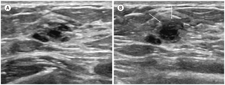
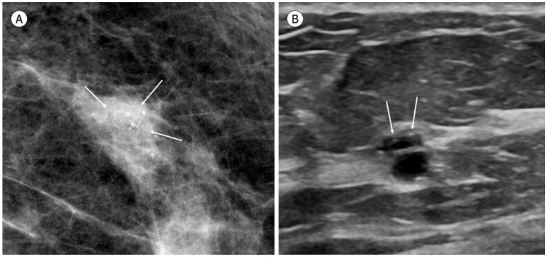
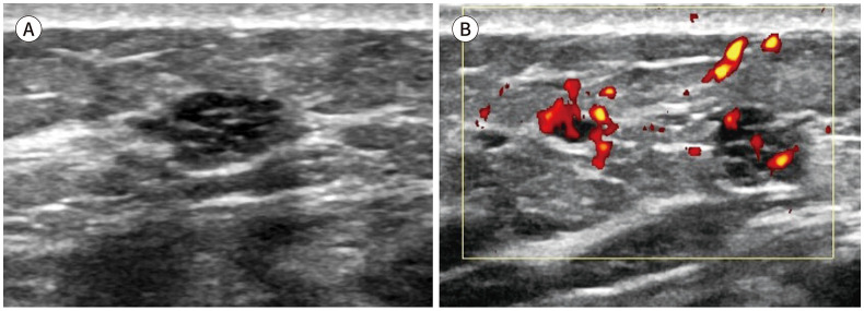
Results: The incidence was 1.5% and 100 patients with 117 lesions were included. Among 117 lesions, 3 (2.6%), 2 (1.7%), and 112 (95.7%) were malignant, high-risk benign, and benign lesions, respectively. The malignant lesions included two cases of ductal carcinoma in situ and one invasive ductal carcinoma. Two of them were assessed as category 4, showing mammographic suspicious microcalcifications and internal vascularity on Doppler US. The remainder was a false negative case and showed echo pattern change on the 12-month follow-up US.
Conclusion: The incidence of clustered microcysts on breast US in asymptomatic women was 1.5% and malignancy rate was 2.6% (3 of 117). Knowledge of outcomes and imaging features of benign and malignant clustered microcysts may be helpful for radiologists, thereby aiding categorization and management recommendations.

 求助内容:
求助内容: 应助结果提醒方式:


